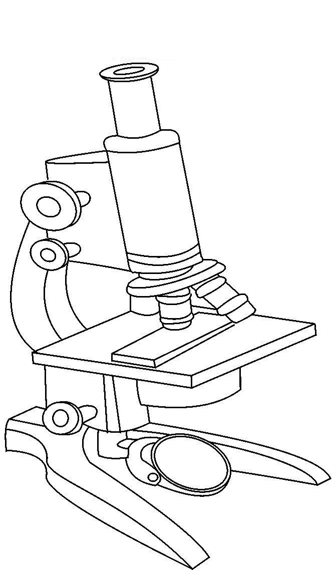
44 picture of compound microscope with label
Science Vocabulary Word List - Enchanted Learning A word list of science vocabulary—from astrophysics to zoology! EnchantedLearning.com is a user-supported site. Site members have full access to an ad-free, print-friendly version of the site. 4.1. Chirality | Organic Chemistry 1: An open textbook In other words, the specific rotation of a chiral compound is the optical rotation that is observed when 1 g of the compound is dissolved in enough of a given solvent to make 100 mL solution, and the rotation is measured in a 1 dm cuvette at 25 o C using light from a sodium lamp.
Virtual Labs: Using the Microscope - GameUp - BrainPOP. In this free online science interactive, students learn the procedures for operating a compound optical light microscope as they would use in a science lab. bVX0-zncj9qJ3G1_r18rkIpQL02X-Oi6tWViR4g4-vwDVmU50WZA-4bRZMjM2TXmc88PAkJ1g0jIembnEbM
Picture of compound microscope with label
Microscope Types (with labeled diagrams) and Functions A compound microscope: Is used to view samples that are not visible to the naked eye Uses two types of lenses - Objective and ocular lenses Has a higher level of magnification - Typically up to 2000x Is used in hospitals and forensic labs by scientists, biologists and researchers to study micro organisms Compound microscope labeled diagram UD Virtual Compound Microscope - University of Delaware ©University of Delaware. This work is licensed under a Creative Commons Attribution-NonCommercial-NoDerivs 2.5 License.Creative Commons Attribution-NonCommercial-NoDerivs 2 Labeling the Parts of the Microscope | Microscope World Resources Labeling the Parts of the Microscope This activity has been designed for use in homes and schools. Each microscope layout (both blank and the version with answers) are available as PDF downloads. You can view a more in-depth review of each part of the microscope here. Download the Label the Parts of the Microscope PDF printable version here.
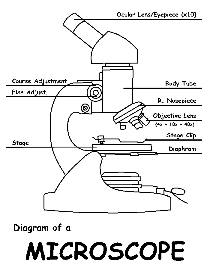
Picture of compound microscope with label. Compound Microscope Labeled Diagram - Quizlet QUESTION. The total magnification of a specimen being viewed with a 10X ocular lens and a 40X objective lens is. 15 answers. QUESTION. a mosquito beats its wings up and down 600 times per second, which you hear as a very annoying 600 Hz sound. if the air outside is 20 C, how far would a sound wave travel between wing beats. 2 answers. Parts of a Compound Microscope - Labeled (with diagrams) A compound microscope is known as a high-power microscope that enables you to achieve a high level of magnification. Smaller specimens can be thoroughly viewed using a compound microscope. Let us take a look at the different parts of a compound microscope and understand each key component. 26+ Picture Of A Microscope With Label PNG 26+ Picture Of A Microscope With Label PNG. Microscopes are specially created to magnify the image of the subject being studied. Students label the parts of the microscope in this photo of a basic laboratory light microscope. Microscope Drawing And Label at GetDrawings | Free download from getdrawings.com I searched for this on bing.com/images. Compound Microscope Parts, Functions, and Labeled Diagram Compound Microscope Definitions for Labels. Eyepiece (ocular lens) with or without Pointer: The part that is looked through at the top of the compound microscope. Eyepieces typically have a magnification between 5x & 30x. Monocular or Binocular Head: Structural support that holds & connects the eyepieces to the objective lenses.
Label the microscope — Science Learning Hub All microscopes share features in common. In this interactive, you can label the different parts of a microscope. Use this with the Microscope parts activity to help students identify and label the main parts of a microscope and then describe their functions. Drag and drop the text labels onto the microscope diagram. Compound Microscope with labels Stock Vector | Adobe Stock Download Compound Microscope with labels Stock Vector and explore similar vectors at Adobe Stock. Adobe Stock. Photos Illustrations Vectors Videos Audio Templates Free Premium Editorial Fonts. ... Get 10 free Adobe Stock images. Start now. Get 10 free images. Unlock 200M+ assets in our full collection. Compound Microscope- Definition, Labeled Diagram, Principle, Parts, Uses In order to ascertain the total magnification when viewing an image with a compound light microscope, take the power of the objective lens which is at 4x, 10x or 40x and multiply it by the power of the eyepiece which is typically 10x. Therefore, a 10x eyepiece used with a 40X objective lens will produce a magnification of 400X. Microscope Parts and Functions Microscope Parts and Functions With Labeled Diagram and Functions How does a Compound Microscope Work?. Before exploring microscope parts and functions, you should probably understand that the compound light microscope is more complicated than just a microscope with more than one lens.. First, the purpose of a microscope is to magnify a small object or to magnify the fine details of a larger ...
Solved Label the image of a compound light microscope using - Chegg Step-by-step answer. Who are the experts? Experts are tested by Chegg as specialists in their subject area. We review their content and use your feedback to keep the quality high. Transcribed image text: Label the image of a compound light microscope using the terms provided. Microscopy - Wikipedia The field of microscopy (optical microscopy) dates back to at least the 17th-century.Earlier microscopes, single lens magnifying glasses with limited magnification, date at least as far back as the wide spread use of lenses in eyeglasses in the 13th century but more advanced compound microscopes first appeared in Europe around 1620 The earliest practitioners of microscopy include Galileo ... Parts of the Microscope with Labeling (also Free Printouts) Parts of the Microscope with Labeling (also Free Printouts) A microscope is one of the invaluable tools in the laboratory setting. It is used to observe things that cannot be seen by the naked eye. Table of Contents 1. Eyepiece 2. Body tube/Head 3. Turret/Nose piece 4. Objective lenses 5. Knobs (fine and coarse) 6. Stage and stage clips 7. Aperture Compound Microscope Stock Photos and Images - Alamy Full-size copy of Robert Hooke's Compound Microscope - held at the Science Museum, London. Hooke, an English natural philosopher, architect and polymath (1635-1703) - author of Micrographia (1665), in which he published results of his microscopic investig Full-size copy of Robert Hooke's Compound Microscope - held at the Science Museum, London.
Microscope Lab - BIOLOGY JUNCTION
A Study of the Microscope and its Functions With a Labeled Diagram To better understand the structure and function of a microscope, we need to take a look at the labeled microscope diagrams of the compound and electron microscope. These diagrams clearly explain the functioning of the microscopes along with their respective parts. ... The camera present within the microscope captures images to reveal the finer ...

parts & components of a light microscope - YouTube
Labeling the Parts of the Microscope | Microscope activity, Science ... Description Worksheet identifying the parts of the compound light microscope. Answer key: 1. Body tube 2. Revolving nosepiece 3. Low power objective 4. Medium power objective 5. High power objective 6. Stage clips 7. Diaphragm 8. Light source 9. Eyepiece 10. Arm 11. Stage 12. Coarse adjustment knob 13. Fine adjustment knob 14. Base
Desenho de Microscópio moderno para colorir - Tudodesenhos
Compound Microscope: Definition, Diagram, Parts, Uses, Working ... - BYJUS Compound microscope is a type of optical microscope that is used for obtaining a high-resolution image. There are more than two lenses in a compound microscope. Learn about the working principle, parts and uses of a compound microscope along with a labeled diagram here.

OMAX Microscope 40X-1000X Student Compound Microscope with Tungsten Light
The flavonoid procyanidin C1 has senotherapeutic activity and ... Dec 06, 2021 · The polyphenol procyanidin C1, a compound found in grape seeds, possesses senomorphic or senolytic activity and is shown to extend the healthspan and survival of old mice and in various models of ...
Free Microscope Drawing, Download Free Microscope Drawing png images ...
Parts of a microscope with functions and labeled diagram Head - This is also known as the body. It carries the optical parts in the upper part of the microscope. Base - It acts as microscopes support. It also carries microscopic illuminators. Arms - This is the part connecting the base and to the head and the eyepiece tube to the base of the microscope.

Parts of the Microscope Printables: Word Searches and More | Life ...
Labelled Diagram of Compound Microscope - Biology Discussion The below mentioned article provides a labelled diagram of compound microscope. Part # 1. The Stand: The stand is made up of a heavy foot which carries a curved inclinable limb or arm bearing the body tube. The foot is generally horse shoe-shaped structure (Fig. 2) which rests on table top or any other surface on which the microscope in kept.
1.5: Microscopy - Biology LibreTexts
A Study of the Microscope and its Functions With a Labeled Diagram ... May 21, 2019 - To better understand the structure and function of a microscope, we need to take a look at the labeled microscope diagrams of the compound and electron microscope. These diagrams clearly explain the functioning of the microscopes along with their respective parts.

Lab 3: Meet the Tetrahymena | CILI CURE Spring 2017
PRACTICAL BOOKLET - BIOLOGY4ISC The first microscope was constructed by Anton Van Leeuwenhoek (1632-1723). This, microscope consisted of a single biconvex lens fitted in a small window of a “board” and the object was viewed through it. This was a simple microscope. After this compound microscope, were developed using combinations of two lenses.

Dog Pancreas Under Microscope Stock Photo - Download Image Now - iStock
Compound Microscope Parts - Labeled Diagram and their Functions - Rs ... Basically, compound microscopes generate magnified images through an aligned pair of the objective lens and the ocular lens. In contrast, "simple microscopes" have only one convex lens and function more like glass magnifiers. [In this figure] Two "antique" microscopes played significant roles in the history of biology.
Post a Comment for "44 picture of compound microscope with label"