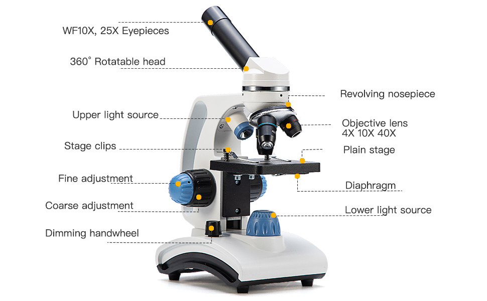
40 drawing of compound microscope with label
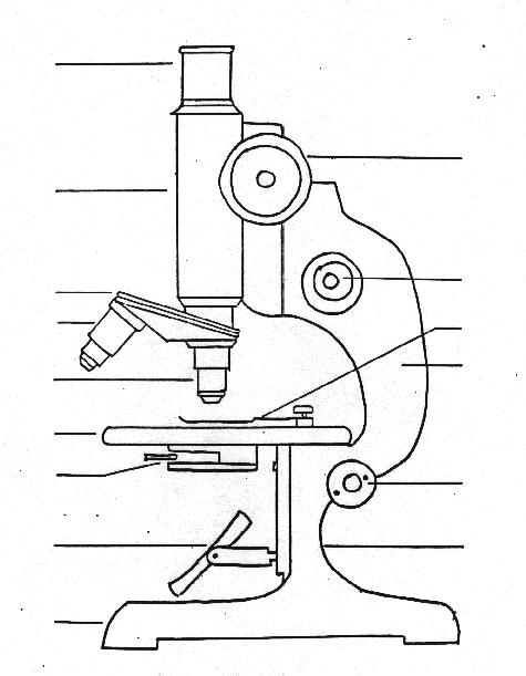
Labelled Diagram of Compound Microscope - Biology Discussion The below mentioned article provides a labelled diagram of compound microscope. Part # 1. The Stand: The stand is made up of a heavy foot which carries a curved inclinable limb or arm bearing the body tube. The foot is generally horse shoe-shaped structure (Fig. 2) which rests on table top or any other surface on which the microscope in kept. Label the microscope - Science Learning Hub Use this interactive to identify and label the main parts of a microscope. Drag and drop the text labels onto the microscope diagram. eye piece lens: The lens you look through - normally 10x or 15x magnification. eye piece lens. coarse focus adjustment: Moves the lens up or down and adjusts focus. coarse focus adjustment.
Compound Microscope- Definition, Labeled Diagram, Principle, Parts, Uses A compound microscope is of great use in pathology labs so as to identify diseases. Various crime cases are detected and solved by drawing out human cells and examining them under the microscope in forensic laboratories. The presence or absence of minerals and the presence of metals can be identified using compound microscopes.

Drawing of compound microscope with label
BIOLOGY 4.docx - Activity 2 Microscopy A. Drawing of Compound ... Activity 2 Microscopy A. Drawing of Compound Microscope with Label B. Give the functions of the following parts of the microscope Parts Functions A. Mechanical Parts Ocular/Eyepiece A small tube consisting of lenses, that indicate the relative power of magnification. Compound Microscope Labeled Diagram - Quizlet QUESTION. The total magnification of a specimen being viewed with a 10X ocular lens and a 40X objective lens is. 15 answers. QUESTION. a mosquito beats its wings up and down 600 times per second, which you hear as a very annoying 600 Hz sound. if the air outside is 20 C, how far would a sound wave travel between wing beats. 2 answers. Label Compound Microscope Lesson Plans & Worksheets The Microscope. For Teachers 5th - 12th. Pupils investigate the parts and functions of a compound microscope. They explore various websites, label the parts of a microscope on a worksheet, view prepared slides, and create drawings of the prepared slides. Get Free Access See Review.
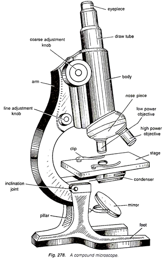
Drawing of compound microscope with label. (i) Draw a neat labelled ray diagram of a compound microscope. Explain ... The eyepiece forms its image A'' B'' which is virtual, erect and magnified. Thus the final image A'' B'' formed by the microscope is inverted and magnified and its position is outside the objective and eyepiece towards objective lens. Magnifying power of compound microscope is. for final image at distance of distinct vision. for final image at ... Compound Microscope Parts, Functions, and Labeled Diagram Compound Microscope Definitions for Labels. Eyepiece (ocular lens) with or without Pointer: The part that is looked through at the top of the compound microscope. Eyepieces typically have a magnification between 5x & 30x. Monocular or Binocular Head: Structural support that holds & connects the eyepieces to the objective lenses. Parts of a microscope with functions and labeled diagram Q. Differentiate between a condenser and an Abbe condenser. Ans. Condensers are lenses that are used to collect and focus light from the illuminator into the specimen. They are found under the stage next to the diaphragm of the microscope. They play a major role in ensuring clear sharp images are produced with a high magnification of 400X and above. Microscope Parts and Functions Most specimens are mounted on slides, flat rectangles of thin glass. The specimen is placed on the glass and a cover slip is placed over the specimen. This allows the slide to be easily inserted or removed from the microscope. It also allows the specimen to be labeled, transported, and stored without damage.
A Study of the Microscope and its Functions With a Labeled Diagram These labeled microscope diagrams and the functions of its various parts, attempt to simplify the microscope for you. However, as the saying goes, 'practice makes perfect', here is a blank compound microscope diagram and blank electron microscope diagram to label. PDF Microscope Practice Actual Size and Drawing Magnification Lab Draw what the object looks like under the microscope. Make sure you get the size of the object compared to the size of the field correct! 3. Label the Name of the object and the Microscope Magnification (Total) beside your drawing. At this point, leave spaces for "Actual Size" & "Drawing Magnification" blank! Draw a ray diagram of compound microscope, when final image is formed ... Draw a ray diagram of compound microscope, when final image is formed at the minimum distance of distinct vision. Easy Solution Verified by Toppr It consist of two convex lenses, one objective of very small focal length with short aperture. And one Eyepiece with moderate focal length and large aperture. Draw a neat labelled diagram of a compound microscope and explain its ... Using sign convention, we find that O'I 1 = + v 0 and O'O = -u where v 0 is the image distance due to the objective and u is the object distance for the objective or the compound microscope. I 1 G 1 is negative and OJ is positive. To find me : The eyepiece behaves like a simple microscope. So : the magnifying power of the eye piece. ∴ m e ...
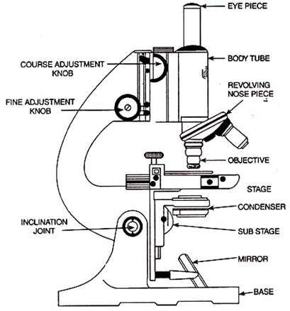
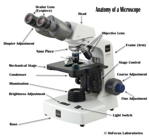
Compound Microscope Parts - Labeled Diagram and their Functions - Rs ... Labeled diagram of a compound microscope Major structural parts of a compound microscope There are three major structural parts of a compound microscope. The head includes the upper part of the microscope, which houses the most critical optical components, and the eyepiece tube of the microscope. Microscope Drawing And Label - Painting Valley microscope diagram compound parts light labeling functions microscopic blank labeled biology microscopy labelled beautiful Compound Microscope ... 496x600 35 0 Parts Of A Compound ... 500x469 27 0 Microscopic Drawing ... 1024x1024 21 4 Download The Diagram... 547x579 17 0 Microscope Labeling ... 270x350 17 0 Microscope Labeling ... 500x529 17 0 draw and label the compound microscope - Brainly.ph Draw and label the compound microscope - 9474237 samanthasolito19 samanthasolito19 19.01.2021 Science Elementary School answered Draw and label the compound microscope 1 See answer Advertisement Advertisement gemjem60 gemjem60 Answer: here I hope this will help. BIOL1200_LabAct2.pdf - Activity 2 Microscopy A. Drawing of Compound ... Compute the total magnification of the Microscope LPO = ( 10x ) ( 10x ) = 100 x HPO = ( 40x ) ( 10x ) =400 x OIO = ( 100x ) ( 10x ) = 1,000 x D. Wet Mounts: Draw the following samples based on the videos presented in the Enrichment task. 1. Letter "e" LPO HPO 2. Cheek Cells (Animal Cell) Unstained LPO HPO Stained LPO HPO 3.
Free Microscope Drawing, Download Free Microscope Drawing png images ...
Compound Microscope Drawing - Painting Valley We collected 37+ Compound Microscope Drawing paintings in our online museum of paintings - PaintingValley.com. ADVERTISEMENT LIMITED OFFER: Get 10 free Shutterstock images - PICK10FREE microscope compound diagram parts functions labeled microscopic binocular art drawing cell huge light labelled unlabeled blank Part Drawing Compoun... 927x1200 58 2

Microscope - The Gemology Project
(a) Draw a labelled ray diagram of compound microscope, when final ... (a) Draw a labelled ray diagram of compound microscope, when final image forms at the least distance of distinct vision. (b) Why is its objective of short focal length and of short aperture, compared to its eyepiece? Explain. (c) The focal length of the objective is 4 cm while that of eyepiece is 10 cm. The object is placed at a distance of 6 cm from the objective lens.
Compound Microscope Drawing With Label - Micropedia
label the parts of the compound microscope - Brainly.ph Activity 2.4: Modern-day Device that Uses Magnetism Directions: •In your notebook, draw an appliance or device that uses magnets. •Below your drawin … g, write a brief explanation of the functions of the magnets present in the device or appliance. •Write the benefits that are provided by the said device or appliance.
Microscope and its types |readbiology.com
How to draw compound of Microscope easily - step by step I will show you " How to draw compound of microscope easily - step by step "Please watch carefully and try this okay.Thanks for watching.....#microscopedrawi...

Microscope | ClipArt ETC
Compound Microscope - Diagram (Parts labelled), Principle and Uses See: Labeled Diagram showing differences between compound and simple microscope parts Structural Components The three structural components include 1. Head This is the upper part of the microscope that houses the optical parts 2. Arm This part connects the head with the base and provides stability to the microscope.
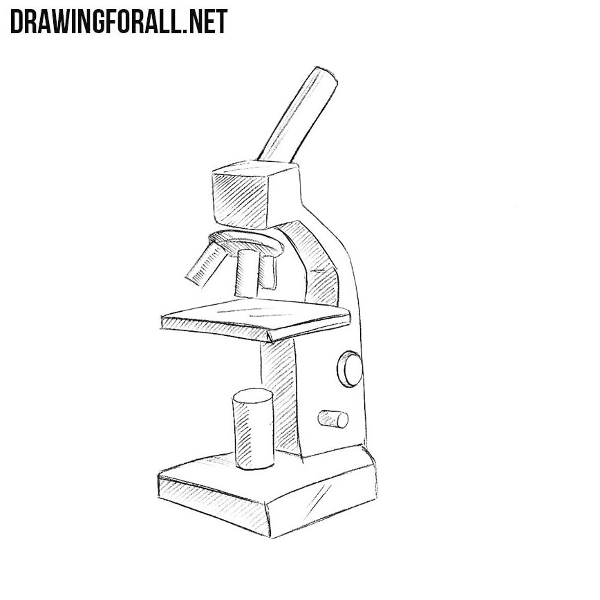
How to Draw a Microscope Easy
BYJUS BYJUS

14 Best Images of Microscope Worksheet Paragraph - Light Microscope ...
(b) Why both objective and eyepiece of a compound microscope must have ... Question (a) Draw the labelled ray diagram for the formation of image by a compound microscope. Derive an expression for its total magnification (or magnifying power), when the final image is formed at the near point. (b) Why both objective and eyepiece of a compound microscope must have short focal lengths?
Compound Microscope Labeled - Micropedia
Microscope Intro - maxineu.bio The Microscope Skills 1. Use a light microscope to investigate the structure of cells and tissues, with drawing of cells 2. Drawing of cell structures as seen with the light microscope. Demonstrate how to draw cell structures seen with a microscope using sharp, carefully drawn lines and straight edge lines for labels.
Lab Flashcards | Easy Notecards
Draw and label a compound microscope? - Answers Draw and label a compound microscope? Wiki User. ∙ 2012-09-19 09:42:29. Study now. See answer (1) Best Answer. Copy. uhmm,.... unang ana is the. 1.) Eyepiece. 2.) arm. eewan ko nakalimutan ko na ...
Post a Comment for "40 drawing of compound microscope with label"