44 light microscope drawing with label
Microscope Drawing: How to Sketch Microscope Slides How to Draw Microscope Slides Organize and orient your field of view: To begin, draw a circle as large as possible with a pencil. An 8.5 x 11-inch piece of paper is good size for beginners. The circle represents what you see through the eyepiece of the microscope. Using thin lines, divide the circle into quarters in order to organize the picture. Labelled Diagram Of A Light Microscope | Products & Suppliers ... Products/Services for Labelled Diagram Of A Light Microscope Microscopes - (706 companies) ...and transmission electron microscopes. Acoustic and ultrasonic microscopes use sound waves to create images of the sample. Compound microscopes use a single light path. These types of microscopes can have a single eyepiece (monocular) or a dual eyepiece...
Microscope Drawing And Label - Painting Valley label microscope diagram compound parts light labeling functions microscopic blank labeled biology microscopy labelled beautiful Compound Microscope ... 496x600 35 0 Parts Of A Compound ... 500x469 27 0 Microscopic Drawing ... 1024x1024 21 4 Download The Diagram... 547x579 17 0 Microscope Labeling ... 270x350 17 0 Microscope Labeling ...
Light microscope drawing with label
Light Microscope: Functions, Parts and How to Use It The function of the light microscope is based on its ability to focus a beam of light through a very small and transparent specimen, to produce an image. The image is then passed through one or two lenses for magnification to view. The transparency of the specimen allows for easy and fast light penetration. Specimens can vary from bacteria to ... How to draw compound of Microscope easily - step by step - YouTube I will show you " How to draw compound of microscope easily - step by step "Please watch carefully and try this okay.Thanks for watching.....#microscopedrawi... PDF The Compound Light Microscope drawing done on blank paper drawing done with sharp pencil firm clear lines (no sketching) no shading/colour used only relevant and easy to see details included large circle drawn to contain drawing labels are neatly printed labels located on right side of drawing labels listed in an even column label lines are parallel and …
Light microscope drawing with label. Microscope Parts and Functions Microscope Parts and Functions With Labeled Diagram and Functions How does a Compound Microscope Work?. Before exploring microscope parts and functions, you should probably understand that the compound light microscope is more complicated than just a microscope with more than one lens.. First, the purpose of a microscope is to magnify a small object or to magnify the fine details of a larger ... Compound Microscope Parts - Labeled Diagram and their Functions - Rs ... The eyepiece (or ocular lens) is the lens part at the top of a microscope that the viewer looks through. The standard eyepiece has a magnification of 10x. You may exchange with an optional eyepiece ranging from 5x - 30x. [In this figure] The structure inside an eyepiece. The current design of the eyepiece is no longer a single convex lens. Light Microscope- Definition, Principle, Types, Parts, Labeled Diagram ... A light microscope is a biology laboratory instrument or tool, that uses visible light to detect and magnify very small objects and enlarge them. They use lenses to focus light on the specimen, magnifying it thus producing an image. The specimen is normally placed close to the microscopic lens. Animal Cell Under Light Microscope Labelled : Draw and label the ... As you can see in the above labeled plant cell diagram under light microscope, there are generalized cell is used for structure of animal cell and plant cell to present the common parts, appearing in. Source: 2.bp.blogspot.com Under a light microscope, the cell membrane, nucleus and cytoplasm of a cheek cell (animal cell) can be observed.
Labeling the Parts of the Microscope | Microscope activity, Science ... Description Worksheet identifying the parts of the compound light microscope. Answer key: 1. Body tube 2. Revolving nosepiece 3. Low power objective 4. Medium power objective 5. High power objective 6. Stage clips 7. Diaphragm 8. Light source 9. Eyepiece 10. Arm 11. Stage 12. Coarse adjustment knob 13. Fine adjustment knob 14. Base 26+ Picture Of A Microscope With Label PNG Microscope pictures with label parts of a microscope with functions and labeled diagram zebra microscope slide labels low price Diagram of microscope with labelling. You must always keep the microscope clean and dry.b. Light or mirror that projects light through the diaphragm. The lenses bend or refract light to make the object beneath them ... Microscope Labeling worksheet Bueno,Emily.docx - WORKSHEET: Compound ... View Microscope Labeling worksheet Bueno,Emily.docx from SCIENCE Biology at Strawberry Crest High School. WORKSHEET: Compound Light Microscope Labeling Instructions: 1. Label the microscope below A Study of the Microscope and its Functions With a Labeled Diagram May 21, 2019 - To better understand the structure and function of a microscope, we need to take a look at the labeled microscope diagrams of the compound and electron microscope. These diagrams clearly explain the functioning of the microscopes along with their respective parts.
Lesson 1.3 The light microscope - Imago Education Draw your own light microscope and label the parts. Use the diagram to show how the microscope works (i.e. add notes to the diagram). Animal cells under a light microscope Learning Activity 2 Answer the following questions in your exercise book. Study Fig. 1.4 on p.3. This shows a generalized animal cell under a light microscope. how to draw microscope (compound) - YouTube drawing microscope. Thank you watching more videos.please subscribe my channel Parts of the Microscope with Labeling (also Free Printouts) Parts of the Microscope with Labeling (also Free Printouts) A microscope is one of the invaluable tools in the laboratory setting. It is used to observe things that cannot be seen by the naked eye. Table of Contents 1. Eyepiece 2. Body tube/Head 3. Turret/Nose piece 4. Objective lenses 5. Knobs (fine and coarse) 6. Stage and stage clips 7. Aperture Label the microscope — Science Learning Hub In this interactive, you can label the different parts of a microscope. Use this with the Microscope parts activity to help students identify and label the main parts of a microscope and then describe their functions. Drag and drop the text labels onto the microscope diagram.
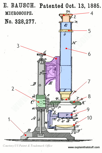
Soft Bone Tissue in a Triceratops Fossil – Proslogion
Label Microscope Diagram - EnchantedLearning.com Using the terms listed below, label the microscope diagram. arm - this attaches the eyepiece and body tube to the base. base - this supports the microscope. body tube - the tube that supports the eyepiece. coarse focus adjustment - a knob that makes large adjustments to the focus. diaphragm - an adjustable opening under the stage, allowing ...
How does a microscope work? - Explain that Stuff
Labeling the Parts of the Microscope Labeling the Parts of the Microscope. This activity has been designed for use in homes and schools. Each microscope layout (both blank and the version with answers) are available as PDF downloads. You can view a more in-depth review of each part of the microscope here.

Microscope Line Art - Free Clip Art
Parts of a microscope with functions and labeled diagram Microscopic illuminator - This is the microscopes light source, located at the base. It is used instead of a mirror. It captures light from an external source of a low voltage of about 100v. Condenser - These are lenses that are used to collect and focus light from the illuminator into the specimen.
![What is a Microscope and How it works [2018]](https://bestmicroscopecentral.com/wp-content/uploads/2017/02/Micrope-Parts-Diagram-Explained.png)
What is a Microscope and How it works [2018]
Microscope Parts, Function, & Labeled Diagram - slidingmotion Condenser. The condenser is to focus the light, which passes from the microscopic illuminator to the specimen. This condenser is located just below the diaphragm. This diaphragm is one of the important parts of the compound microscope which will help to get an accurate and sharp image. The condenser has a magnification power of 400X and above.
Sample Descriptive Lab Report
Light Microscope Drawing - Painting Valley light microscope parts compound diagram microscopy label schematic electron lab blank appliances measurement sampling optical between working microscopic labeled functions polarized Diagram Of A Compoun... 414x467 4 0 Part Drawing Light M... 927x1200 2 0 Compound Light Micro... 630x380 2 0 Applications And Ski... 776x461 1 2 Microscopic Drawing ...

Microscope With Labels Clip Art at Clker.com - vector clip art online ...
Microscope, Microscope Parts, Labeled Diagram, and Functions Illuminator: Illuminator is the most important microscope parts and it serve as light source for a microscope during slide specimen visualization. It is a continuous source of light (110 volts) used in place of a mirror. The mirror of microscope is used to reflect light from the external light source up through the bottom of the stage.
Biology label part of microscope
Compound Light Microscope Labeling - Printable - PurposeGames.com About this Worksheet. This is a free printable worksheet in PDF format and holds a printable version of the quiz Compound Light Microscope Labeling.By printing out this quiz and taking it with pen and paper creates for a good variation to only playing it online.
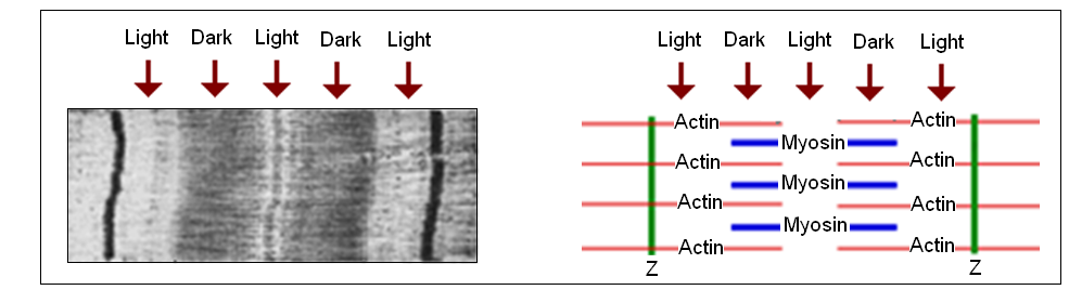
Topic 11.2: Movement - AMAZING WORLD OF SCIENCE WITH MR. GREEN
How to Sketch a Microscope Slide - Identifying and Sketching Cell ... For a pencil sketch, separate areas into white, light, medium and dark grey and black. To see the light/dark areas, squint so that the hard edges are blurred and your focus is on the shading. Start shading the light areas by following the shapes. For example, shade vertical lines for a flat surface and curved lines for a rounded.
Post a Comment for "44 light microscope drawing with label"