40 phospholipid drawing labeled
The Lipid Bilayer - Molecular Biology of the Cell - NCBI Bookshelf The lipid bilayer has been firmly established as the universal basis for cell-membrane structure. It is easily seen by electron microscopy, although specialized techniques, such as x-ray diffraction and freeze-fracture electron microscopy, are needed to reveal the details of its organization. The bilayer structure is attributable to the special properties of the lipid molecules, which cause ... Phospholipid Bilayer | Lipid Bilayer | Structures & Functions As portrayed in the diagrammatic illustration above, the glycerol molecule and the phosphate group makes up the " hydrophilic " head, or the water-loving part of the phospholipid. This head is such because of the negatively charged phosphate group that tends to attract the water molecules.
quizlet.com › 590125929 › biochemistry-final-reviewBiochemistry Final Review Questions Flashcards | Quizlet The plasma membrane of E. coli is approximately 75% protein and 25% phospholipid by weight. It is known that the average membrane protein molecular weight is 50,000 Da and an average membrane phospholipid molecular weight is 750 Da. Calculate the number of membrane phospholipid molecules present per molecule of membrane protein. CH 11 Q 10

Phospholipid drawing labeled
› books › NBK26871The Lipid Bilayer - Molecular Biology of the Cell - NCBI ... The motion and orientation of a spin-labeled lipid in a bilayer can be deduced from the ESR spectrum. Such studies show that phospholipid molecules in synthetic bilayers very rarely migrate from the monolayer (also called a leaflet) on one side to that on the other. This process, known as “flip-flop,” occurs less than once a month for any ... Draw and Label a Phospholipid | Lipids | A Level Biology This short clip from the Lesson "Lipids: - the properties of Phospholipids", You'll learn the structure of a phospholipid - which is a commonly expected "id... Phospholipid Structure Labeling Diagram | Quizlet Start studying Phospholipid Structure Labeling. Learn vocabulary, terms, and more with flashcards, games, and other study tools. Start a free trial of Quizlet Plus by Thanksgiving | Lock in 50% off all year Try it free
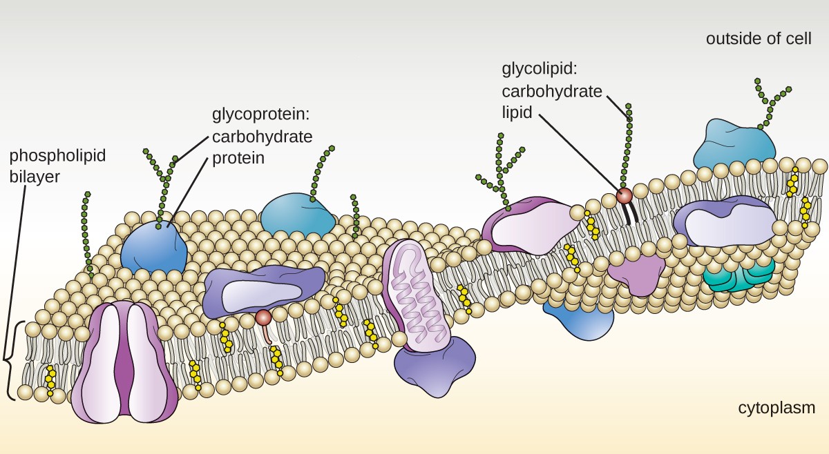
Phospholipid drawing labeled. Label the Phospholipid Bilayer Diagram | Quizlet Only $35.99/year Label the Phospholipid Bilayer STUDY Learn Flashcards Write Spell Test PLAY Match Gravity Created by Ava_Amici Terms in this set (8) phospholipid composed of a hydrophobic tail and a hydrophilic head hydrophilic heads Negative charge so they attract to water hydrophobic tails Fatty acids are nonpolar and hydrophobic cholesterol Structure of Phospholipids (With Diagram) | Lipid Metabolism 1. Phosphatidylcholine (Lecithin). This phospholipid has nitrogen containing choline in its phosphorylated component. 2. Phosphatidylethanolamine (Cephalin). The phosphorylated component contains ethanolamine here. ADVERTISEMENTS: 3. Phosphatidylinositol. This phospholipid contains hexahydric alcohol called inositol in its phosphorylated component. File:Cell membrane detailed diagram 4.svg - Wikipedia Cell membrane detailed diagram 4.svg. English: The cell membrane, also called the plasma membrane or plasmalemma, is a semipermeable lipid bilayer common to all living cells. It contains a variety of biological molecules, primarily proteins and lipids, which are involved in a vast array of cellular processes. Phospholipids | Introduction to Chemistry | | Course Hero Phospholipid Molecule A phospholipid is a molecule with two fatty acids and a modified phosphate group attached to a glycerol backbone. The phosphate may be modified by the addition of charged or polar chemical groups. Two chemical groups that may modify the phosphate, choline and serine, are shown here.
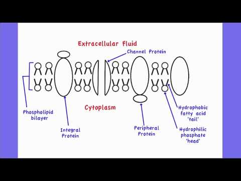
Phospholipid Vector & Photo (Free Trial) | Bigstock Download high-quality Phospholipid phosphatides lipids microscopical images, illustrations and vectors perfectly priced to fit your projects budget. ... Phospholipid or phosphatides lipids microscopical structure outline diagram. Labeled educational description with cells hydrophilic head, hydrophobic tail and extracellular space vector ... Lipids - Michigan State University The phospholipid molecules can move about in their half the bilayer, but there is a significant energy barrier preventing migration to the other side of the bilayer. To see an enlarged segment of a phospholipid bilayer Click Here. This bilayer membrane structure is also found in aggregate structures called liposomes. Liposomes are microscopic ... › articles › s41573/020/0073-9Radiopharmaceutical therapy in cancer: clinical ... - Nature Jul 29, 2020 · RPT has the benefit of drawing on the substantial knowledge base of ... Phospholipid ether analogues. ... evaluation of 125 I- vs. 131 I-labeled CO17-1A in a human colorectal cancer ... 2.4.1 Draw and label a diagram to show the structure of membranes when drawing and labeling a diagram of the plasma membrane you should be sure to include: the phospholipid bilayer with hydrophobic 'tails' and hydrophilic 'heads' of the phospholipids labelled...
Biochemistry Final Review Questions Flashcards | Quizlet The plasma membrane of E. coli is approximately 75% protein and 25% phospholipid by weight. It is known that the average membrane protein molecular weight is 50,000 Da and an average membrane phospholipid molecular weight is 750 Da. Calculate the number of membrane phospholipid molecules present per molecule of membrane protein. CH 11 Q 10 Phospholipid Bilayer | Introduction, Structure and Functions Phospholipid Diagram. Phospholipid Structure. A Phospholipid molecule is comprised of two Fatty Acid tails and Phosphate Group which make its Head. Fatty acids are chemically composed of long chains of Hydrogen and Carbon atoms. While Phosphate groups comprised of a Phosphorus molecule. Four oxygen molecules attached to Phosphate group. Draw And Label A Phospholipid Bilayer : Phospholipids Images Stock ... The phospholipid molecule draw and label this: Biological membranes usually involve two layers of phospholipids with their tails pointing inward, an arrangement called a phospholipid bilayer. Remember that the cell membrane is a lipid bi layer in this lipid . When drawing and labeling a diagram of the plasma membrane you should be sure to ... Bird Respiratory System - EKU Tools Semi-schematic drawing of the lung-air sac system in situ. ... SP-A, and (phospholipid) regulators of inflammatory processes (From: Bernhard et al. 2004). A: A high-power view of a foreign particle (p) being engulfed by an epithelial cell (e) in an avian lung. ... labeled with the presence of erythrocytes (*), and air capillaries (AC) that make ...
What Is The Makeup Of The Cell Membrane | Saubhaya Makeup
Phospholipid - Wikipedia Phospholipids, are a class of lipids whose molecule has a hydrophilic "head" containing a phosphate group and two hydrophobic "tails" derived from fatty acids, joined by an alcohol residue (usually a glycerol molecule). Marine phospholipids typically have omega-3 fatty acids EPA and DHA integrated as part of the phospholipid molecule.

Cell Theory, Form, and Function: Fluid Mosaic Model of Membrane ...
› articles › s41467/022/30300-zTMEM16 scramblases thin the membrane to enable lipid ... - Nature May 11, 2022 · Briefly lipids in chloroform (Avanti), including 0.4% w/w tail labeled NBD-PE, were dried under N 2, washed with pentane and resuspended at 20 mg ml −1 in 150 mM KCl, 50 mM HEPES pH 7.4 with 35 ...
1. Neurobiology
Phospholipid structure (video) | Cells | Khan Academy Video transcript. In this video, we're going to actually explore in detail the structure of phospholipids in our cell membrane. Just to briefly remind us, our phospholipid is often drawn like this. It has that polar phosphate head group, and it has two fatty acid chains. And all of this is held together by glycerol backbone.
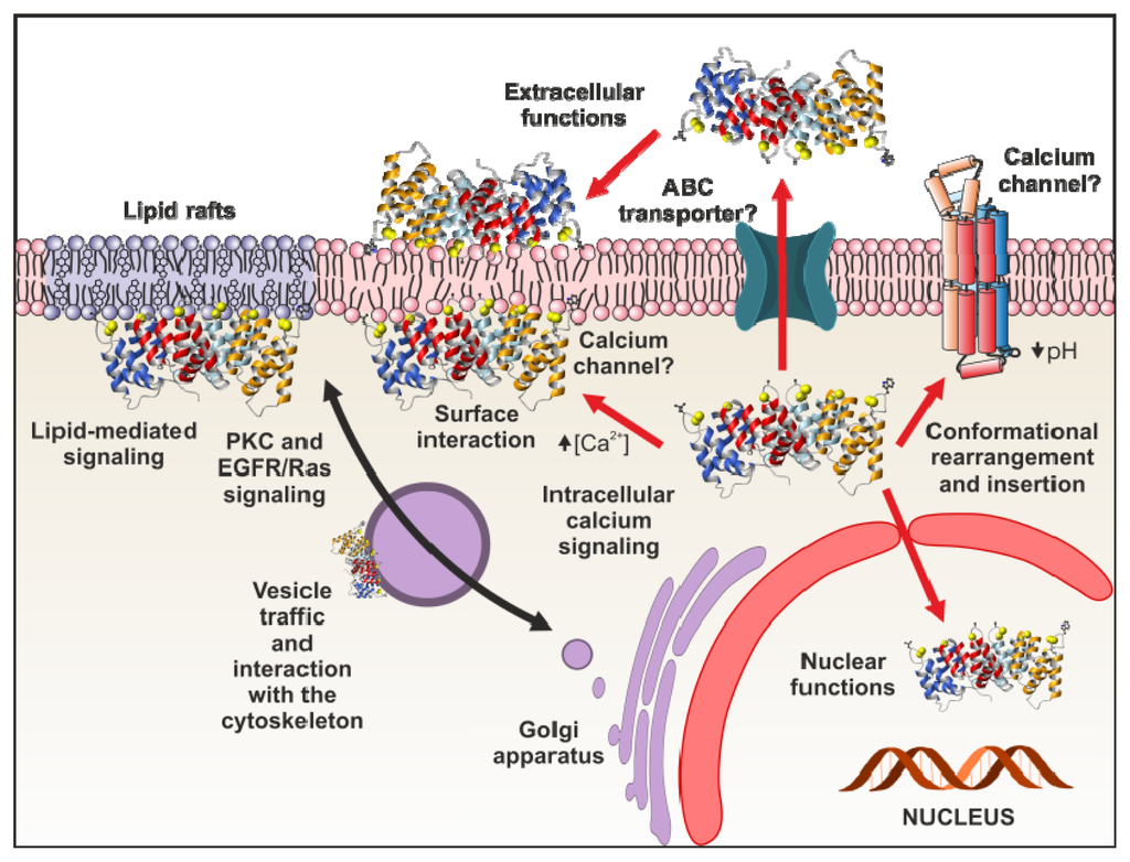
Annexine A6; Annexine VI; Calcimédine 67 kDa; Calélectrine 67 kDa ...
Solved Drawing #1: Phospholipid Bilayer a Draw a labeled | Chegg.com Transcribed image text: Drawing #1: Phospholipid Bilayer a Draw a labeled diagram that shows how 10 molecules of phospholipid would naturally arrange themselves if they were dropped into a cup of water. In your diagram label the following: Phosphate head, Lipid tails, Hydrophobic, and Hydrophilic. Drawing #2: Water a Draw a labeled diagram that shows how 3 molecules of water would naturally ...
2.4.1 Draw and label a diagram to show the structure of membranes - YouTube
TMEM16 scramblases thin the membrane to enable lipid ... - Nature May 11, 2022 · Briefly lipids in chloroform (Avanti), including 0.4% w/w tail labeled NBD-PE, were dried under N 2, washed with pentane and resuspended at 20 mg ml −1 in 150 mM KCl, 50 mM HEPES pH 7.4 with 35 ...

B for Biology: Cell Membrane - Protector of the Cell
Phospholipid Bilayer Cell Membrane Diagram Labeled / The Fluid Mosaic ... The plasma membrane is composed of a phospholipid bilayer. Structure of the cell membrane; The plasma membrane structure as a mosaic of phospholipids, cholesterol, . These consist of a head molecule, a phosphate . The membrane bilayer contains many kinds of phospholipid molecules, with different sized head and tail molecules.
Unique Characteristics of Eukaryotic Cells | Microbiology
Phospholipids - Structure, Types, Properties and Function A phospholipid is a molecule containing a glycerol backbone and two fatty acids linked, as well as a modified phosphate group. The addition of charged or polar chemical groups to the phosphate can change its properties. Choline and serine, two chemical groups that can alter phosphate.
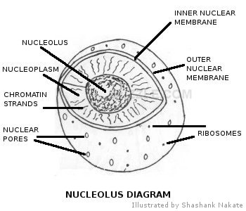
The Control Center - Cells & Organelles
Organic & Biomolecular Chemistry Ruthenium photoredox-triggered phospholipid membrane formation From DOI: 10.1039/C6OB00290K. ... Following the preparation of Cy7.5-labeled AGE-albumin and intravenous injection in BALB/cA-nu/nu mice, noninvasive fluorescence kinetics analysis was performed. ... When drawing structures or schemes, authors should use the correct sizes and ...
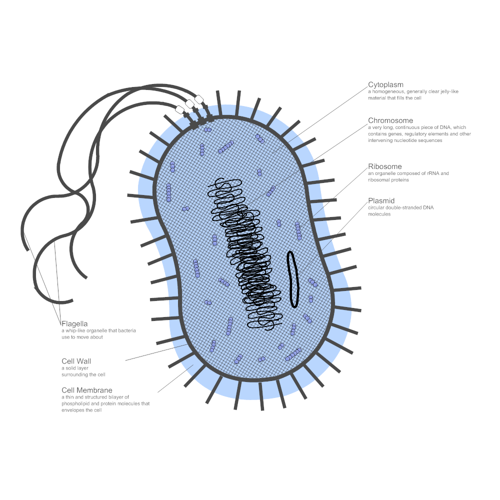
Bacteria Diagram
Solved Drawing #1: Phospholipid Bilayer Draw a labeled | Chegg.com Drawing #1: Phospholipid Bilayer Draw a labeled diagram that shows how 10 molecules of phospholipid would naturally arrange themselves if they were dropped into a cup of water. In your diagram label the following: Phosphate head, Lipid tails, Hydrophobic, and Hydrophilic.
Post a Comment for "40 phospholipid drawing labeled"