41 labelled diagram of compound microscope
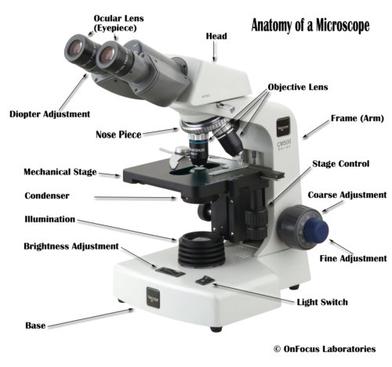
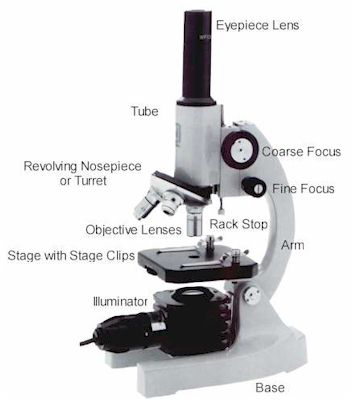
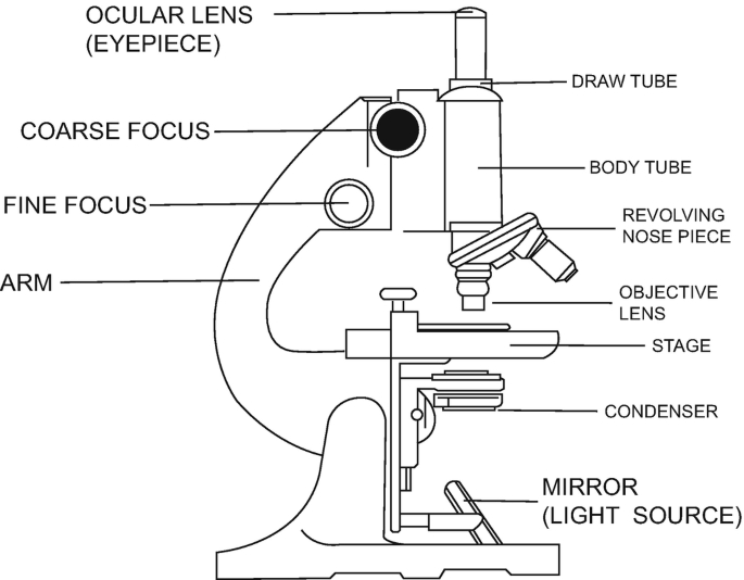
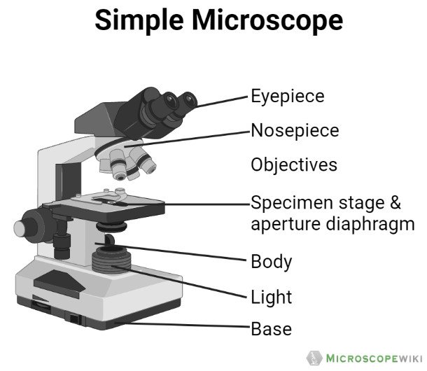
Parts of the Microscope (Labeled Diagrams) - Simple and Compound Microscope It holds the microscope upright. Simple microscope labelled diagram Image created with Biorender Tube/Body Tube It serves as the connector between the eyepiece/ocular and objective lenses. Objective lenses The lenses have varying magnifying power, which typically consists of 10x, 40x, and 100x. Draw a neat labelled diagram of a compound microscope ... - Vedantu The Optical Parts of Compound Microscope include: 1. Eyepiece lens or Ocular: At the top of the body tube, a lens is planted which is known as the eyepiece. On ...
Compound microscope - their parts and function - Microscopy4kids Labeled diagram of a compound microscope. Optical components of a compound microscope. The term "compound" refers to the microscope having more than one lens. Compound microscopes generate magnified images through an aligned pair of the objective lens and the ocular lens. In contrast, "simple microscopes" have only one convex lens and ...

Labelled diagram of compound microscope
Compound Microscope – Diagram (Parts labelled), Principle and ... Oct 10, 2022 · Compound Microscope Parts (Labeled diagram) A compound microscope basically consists of optical and structural components. Within these two systems, there are multiple components within them and they are: Image : Labeled Diagram of compound microscope parts See: Labeled Diagram showing differences between compound and simple microscope parts how to Draw Compound Microscope step by step, Labelled Diagram Jan 18, 2023 ... how to Draw Compound Microscope step by step, Labelled Diagram#compoundmicroscope #diagram #biologydiagram #howtodraw. A Study of the Microscope and its Functions With a Labeled Diagram ... These labeled microscope diagrams and the functions of its various parts, attempt to simplify the microscope for you. However, as the saying goes, 'practice makes perfect', here is a blank compound microscope diagram and blank electron microscope diagram to label. Download the diagrams and practice labeling the different parts of these ...
Labelled diagram of compound microscope. Compound Microscope- Definition, Labeled Diagram, Principle, Parts, Uses A compound light microscope mostly uses a low voltage bulb as an illuminator. The stage is the flat platform where the slide is placed. Nosepiece and Aperture Nosepiece is a rotating turret that holds the objective lenses. The viewer spins the nosepiece to select different objective lenses. Microscope Diagram Labeled, Unlabeled and Blank | Parts of a Microscope Microscope Diagram Labeled First and foremost, we have a labeled microscope diagram, available in both black and white and color. Useful as a study guide for learning the anatomy of a microscope. There are six printables available. Compound Microscope Principle, Parts, Diagram Definition, Application Using two sets of lenses, a compound microscope amplifies the image under the microscope. The compound microscope contains multiple convex lenses. It is equipped with a 4x, 10x, 40x, and 100x objective lens and a 10x eyepiece. Compound microscopes are used to view small samples that cannot be seen by the naked eye. Diagram of a Compound Microscope - Biology Discussion Diagram of a Compound Microscope Article Shared by ADVERTISEMENTS: In this article we will discuss about:- 1. Essential Parts of Compound Microscope 2. Magnification of the Image of the Object by Compound Microscope 3. Resolution Power 4. Method for Studying Microbes 5. Measurement of the Size of Objects. Essential Parts of Compound Microscope:
Parts of a Compound Microscope - Labeled (with diagrams) Parts of a Compound Microscope - Labeled (with diagrams) A compound microscope is known as a high-power microscope that enables you to achieve a high level of magnification. Smaller specimens can be thoroughly viewed using a compound microscope. Let us take a look at the different parts of a compound microscope and understand each key component. Microscope Parts and Functions With Labeled Diagram and ... Microscope Parts and Functions With Labeled Diagram and Functions How does a Compound Microscope Work?. Before exploring microscope parts and functions, you should probably understand that the compound light microscope is more complicated than just a microscope with more than one lens.. First, the purpose of a microscope is to magnify a small object or to magnify the fine details of a larger ... Draw a labelled ray diagram of an image formed by a compound ... Draw a labelled ray diagram of an image formed by a compound microscope, when the final image lies at the least distance of distinct vision (D). Draw a neat labelled diagram of a compound microscope and ... Apr 26, 2023 ... Description : It consists of two convex lenses separated by a distance. The lens near the object is called objective and the lens near the ...
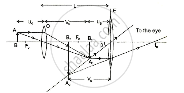
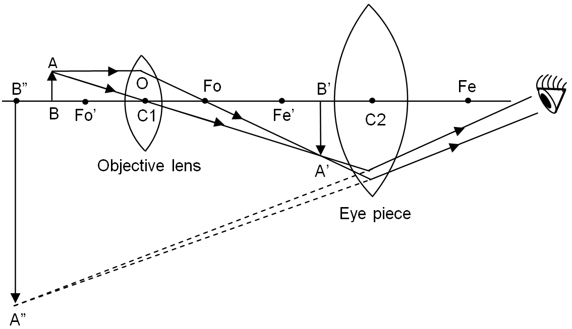
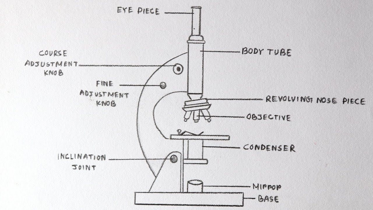
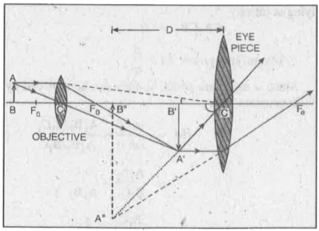
16 Parts of a Compound Microscope: Diagrams and Video The 16 core parts of a compound microscope are: Head (Body) Arm Base Eyepiece Eyepiece tube Objective lenses Revolving Nosepiece (Turret) Rack stop Coarse adjustment knobs Fine adjustment knobs Stage Stage clips Aperture Illuminator Condenser Diaphragm Video: Parts of a compound Microscope with Diagram Explained Compound Microscope Parts, Functions, and Labeled Diagram Compound Microscope Definitions for Labels Eyepiece (ocular lens) with or without Pointer: The part that is looked through at the top of the compound microscope. Eyepieces typically have a magnification between 5x & 30x. Monocular or Binocular Head: Structural support that holds & connects the eyepieces to the objective lenses. Draw a labelled ray diagram of a compound microscope and ... - Vedantu In this case, the objective lens O of the compound microscope forms a real, inverted and enlarged image A'B' of the object. Now A'B' acts as an object for the eyepiece E, whose position is adjusted so that A'B' lies between optical centre C2 and the focus fe' of eyepiece. Now the eyepiece forms a final virtual, inverted and highly ... Parts of a microscope with functions and labeled diagram - Microbe Notes There are three structural parts of the microscope i.e. head, base, and arm. Head - This is also known as the body. It carries the optical parts in the upper part of the microscope. Base - It acts as microscopes support. It also carries microscopic illuminators.
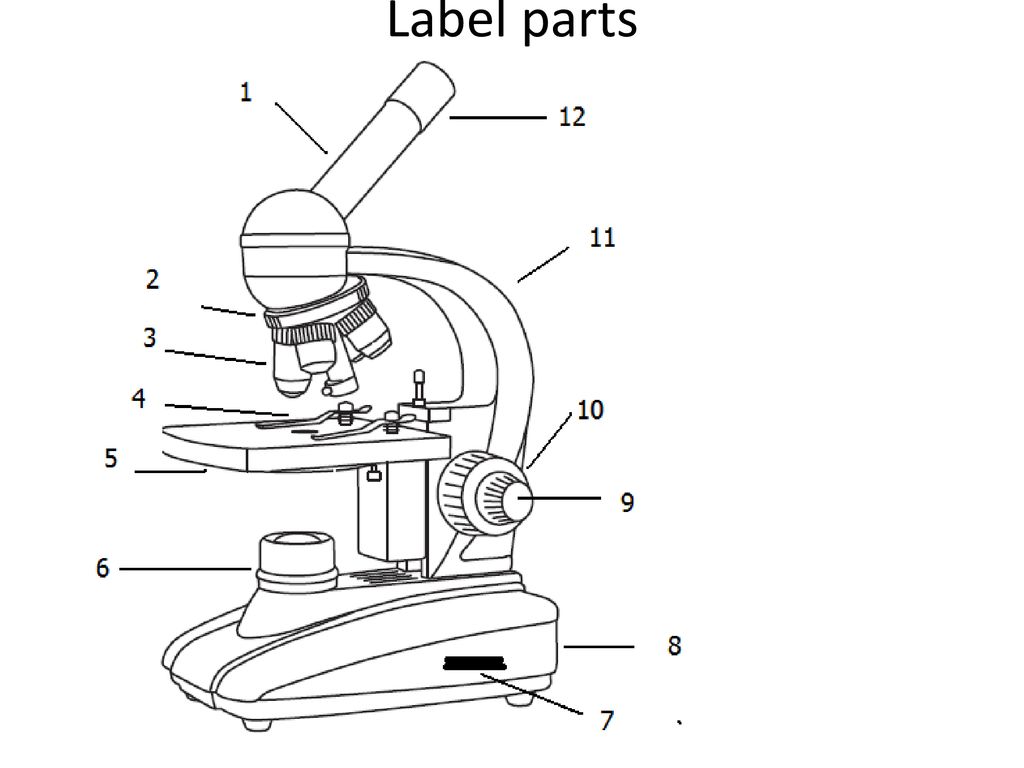
Labeling the Parts of the Microscope | Microscope World Resources
(i) Draw a neat labelled ray diagram of a compound microscope ... May 15, 2018 ... diagram of a compound microscope. Working: Suppose a small object AB is placed slightly away from the first focus F0' of the objective lens.
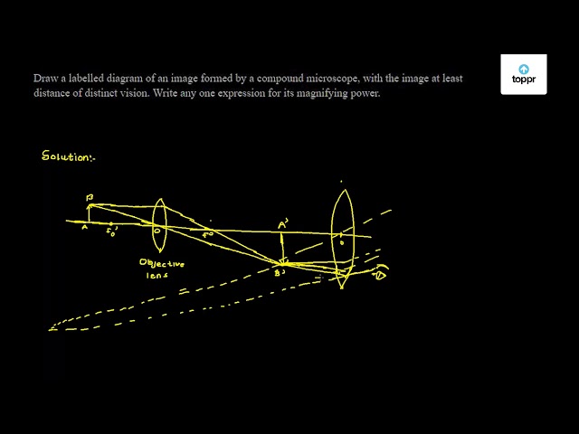
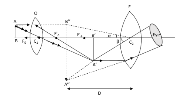
Draw a labelled diagram of an image formed by a compound microscope, with the image at least distance of distinct vision. Write any one expression for its magnifying power.
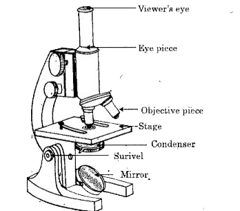
Labelled Diagram of Compound Microscope - Biology Discussion The below mentioned article provides a labelled diagram of compound microscope. Part # 1. The Stand: The stand is made up of a heavy foot which carries a curved inclinable limb or arm bearing the body tube. The foot is generally horse shoe-shaped structure (Fig. 2) which rests on table top or any other surface on which the microscope in kept.

Draw a well labelled diagram of a microscope. - Brainly.in
Compound Microscope Parts – Labeled Diagram and their ... There are three major structural parts of a compound microscope. The head includes the upper part of the microscope, which houses the most critical optical components, and the eyepiece tube of the microscope. The base acts as the foundation of microscopes and houses the illuminator. The arm connects between the base and the head parts.

easy compound microscope diagram - Clip Art Library
Compound Microscope Parts, Function, & Diagram - Study.com Learn the compound light microscope's parts and functions by viewing a compound microscope diagram. Also, read about the uses of a compound microscope. Updated: 11/04/2021
Microscope Parts and Functions
Microscopy: Intro to microscopes & how they work (article) - Khan Academy In most cases, the part of a cell or tissue that we want to look at isn't naturally fluorescent, and instead must be labeled with a fluorescent dye or tag before it goes on the microscope. The leaf picture at the start of the article was taken using a specialized kind of fluorescence microscopy called confocal microscopy.
Solved Nikon Parts of the compound microscope Write the ...
Draw a labelled diagram of an image formed by a compound ... - Toppr Expression of magnifying power of a compound microscope is given by: m=−uovo(1+feD) Where vo is the image distance from the objective lens

Optical Instruments - Compound Microscope - Basic ...
Compound Microscope: Definition, Diagram, Parts, Uses ... The compound microscope is mainly used for studying the structural details of cell, tissue, or sections of organs. The parts of a compound microscope can be classified into two: Non-optical parts Optical parts Non-optical parts Base The base is also known as the foot which is either U or horseshoe-shaped.

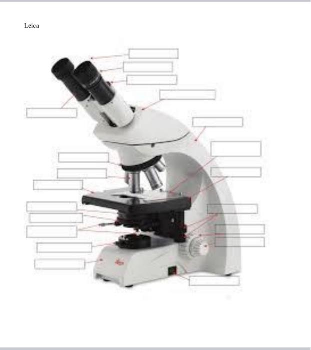
Microscope Labeling
Label the microscope — Science Learning Hub All microscopes share features in common. In this interactive, you can label the different parts of a microscope. Use this with the Microscope parts activity to help students identify and label the main parts of a microscope and then describe their functions.. Drag and drop the text labels onto the microscope diagram. If you want to redo an answer, click on the box and the answer will go back ...

Draw a neat labelled diagram of a compound microscope. Derive ...
Compound Microscope Labeled Diagram | Quizlet Compound Microscope Labeled + − Flashcards Learn Test Match Created by meganplocher734 Terms in this set (14) Eyepiece/Ocular lens Contains the ocular lens Body tube A hollow cylinder that holds the eyepiece. Arm Part that supports the microscope. Stage Supports the slide or specimen Coarse adjustment Knob
Draw a Labelled Ray Diagram Showing the Formation of a Final ...
Compound Microscope Labeled Definition, Labeled Diagram, Best procedure ... A compound microscope or compound microscope labeled is a type of microscope that uses two or more lenses to magnify an object. The first lens, called the eyepiece, is used to view the object. The second lens, called the objective lens, is used to collect light from the object and focus it on the eyepiece.

Compound Microscope Labeling Part 1 Diagram | Quizlet
A Study of the Microscope and its Functions With a Labeled Diagram ... These labeled microscope diagrams and the functions of its various parts, attempt to simplify the microscope for you. However, as the saying goes, 'practice makes perfect', here is a blank compound microscope diagram and blank electron microscope diagram to label. Download the diagrams and practice labeling the different parts of these ...
Draw a neat labelled diagram of a compound microscope and ...
how to Draw Compound Microscope step by step, Labelled Diagram Jan 18, 2023 ... how to Draw Compound Microscope step by step, Labelled Diagram#compoundmicroscope #diagram #biologydiagram #howtodraw.
a) Draw a labelled ray diagram of a compound microscope. (b ...
Compound Microscope – Diagram (Parts labelled), Principle and ... Oct 10, 2022 · Compound Microscope Parts (Labeled diagram) A compound microscope basically consists of optical and structural components. Within these two systems, there are multiple components within them and they are: Image : Labeled Diagram of compound microscope parts See: Labeled Diagram showing differences between compound and simple microscope parts

Label the Microscope Diagram | Download Scientific Diagram
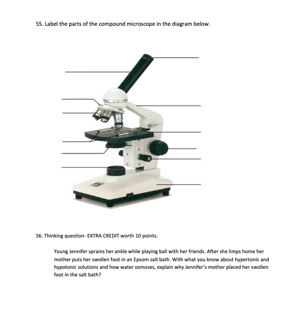
Solved 55. Label the parts of the compound microscope in the ...

Compound Microscope Parts, Functions, and Labeled Diagram ...
Microscope Parts & Specifications Labeled Diagram ...

Draw a neat labelled diagram of a compound microscope and ...
Exercise 1: Using a Compound Microscope | SpringerLink

Simple Microscope Definition, Magnification, Parts And Uses
Compound Microscope Review - ppt download
Draw a labelled ray diagram of a compound microscope and ...
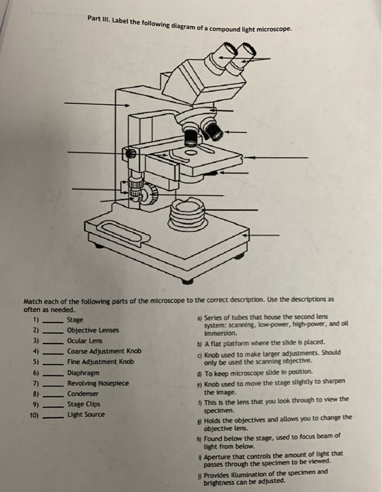
Solved Part III. Label the following diagram of a compound ...
Draw a labelled ray diagram of an image formed by a compound ...

Cytology. Cytology. radiation used to illuminate the specimen ...

Compound Microscope Parts – Labeled Diagram and their ...
Parts of the Microscope (Labeled Diagrams) - Simple and ...

How to draw compound of Microscope easily - step by step

Compound Microscope Principle, Parts, Diagram Definition ...

Compound Microscope Parts, Functions, and Labeled Diagram ...
Label microscope - Teaching resources

File:Labelledmicroscope.gif - Wikibooks, open books for an ...
Draw a labelled diagram of a compound microscope.

Draw a labelled ray diagram of compound microscope and derive ...

Labelled Diagram of Compound Microscope | Figure Of Compound ...
How to draw Compound of Microscope easily - step by step | How to draw Microscope diagram
Draw a labelled diagram of compound microscope. Derive ...

Label the microscope — Science Learning Hub

give a well labelled diagram of compound microscope using of ...
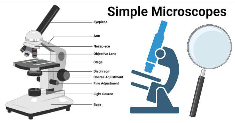
Simple Microscope- Definition, Principle, Magnification ...

Microscope With Labels Clip Art at Clker.com - vector clip ...
Post a Comment for "41 labelled diagram of compound microscope"