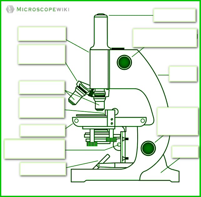
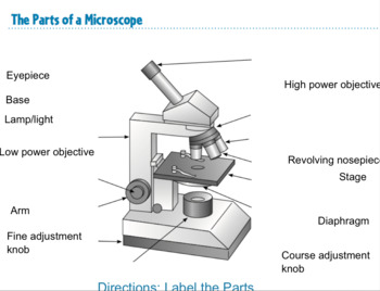
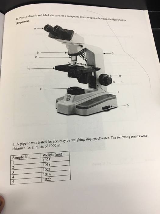
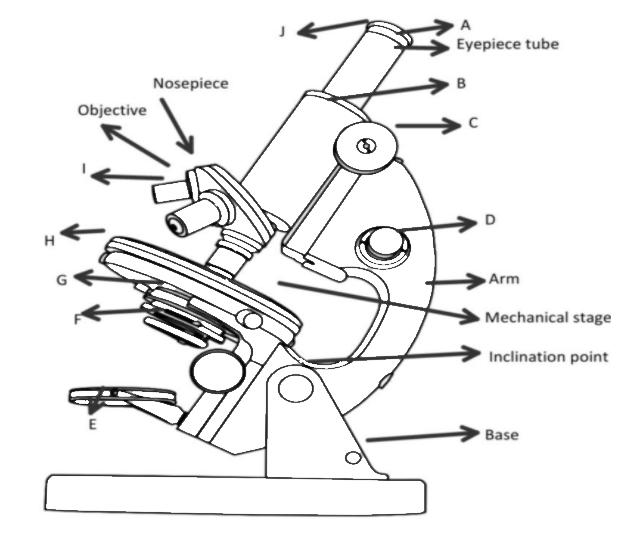
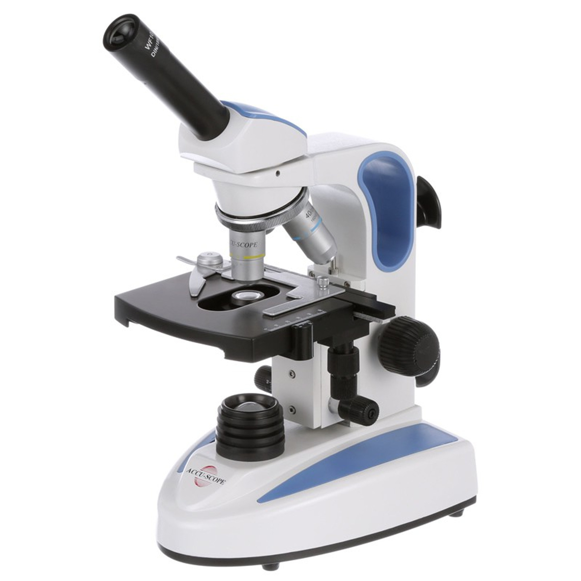
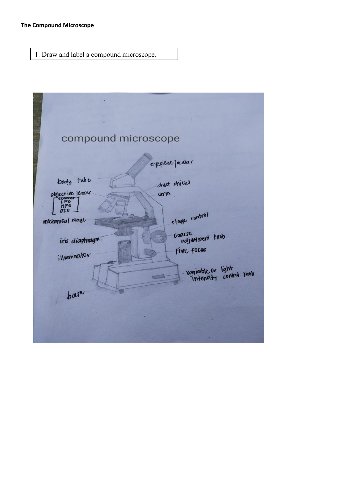
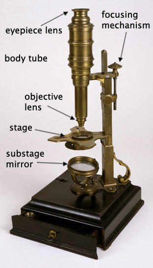
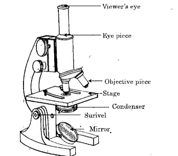
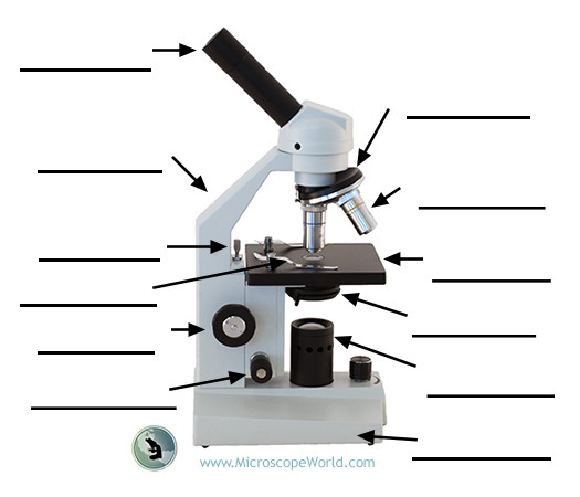
40 labelled compound microscope
How to draw compound of Microscope easily - step by step I will show you " How to draw compound of microscope easily - step by step "Please watch carefully and try this okay.Thanks for watching.....#microscopedrawi... Parts of a Microscope - SmartSchool Systems Labeled Parts of a Microscope Word Document. Labeled Parts of a Microscope PDF. Unlabeled Microscope Parts Worksheets. Image - JPG. Word Document. PDF. ... Most compound microscopes come with three or four objective lenses that revolve on the nosepiece. The most common objective lenses have power of 4X, 10X and 40X.
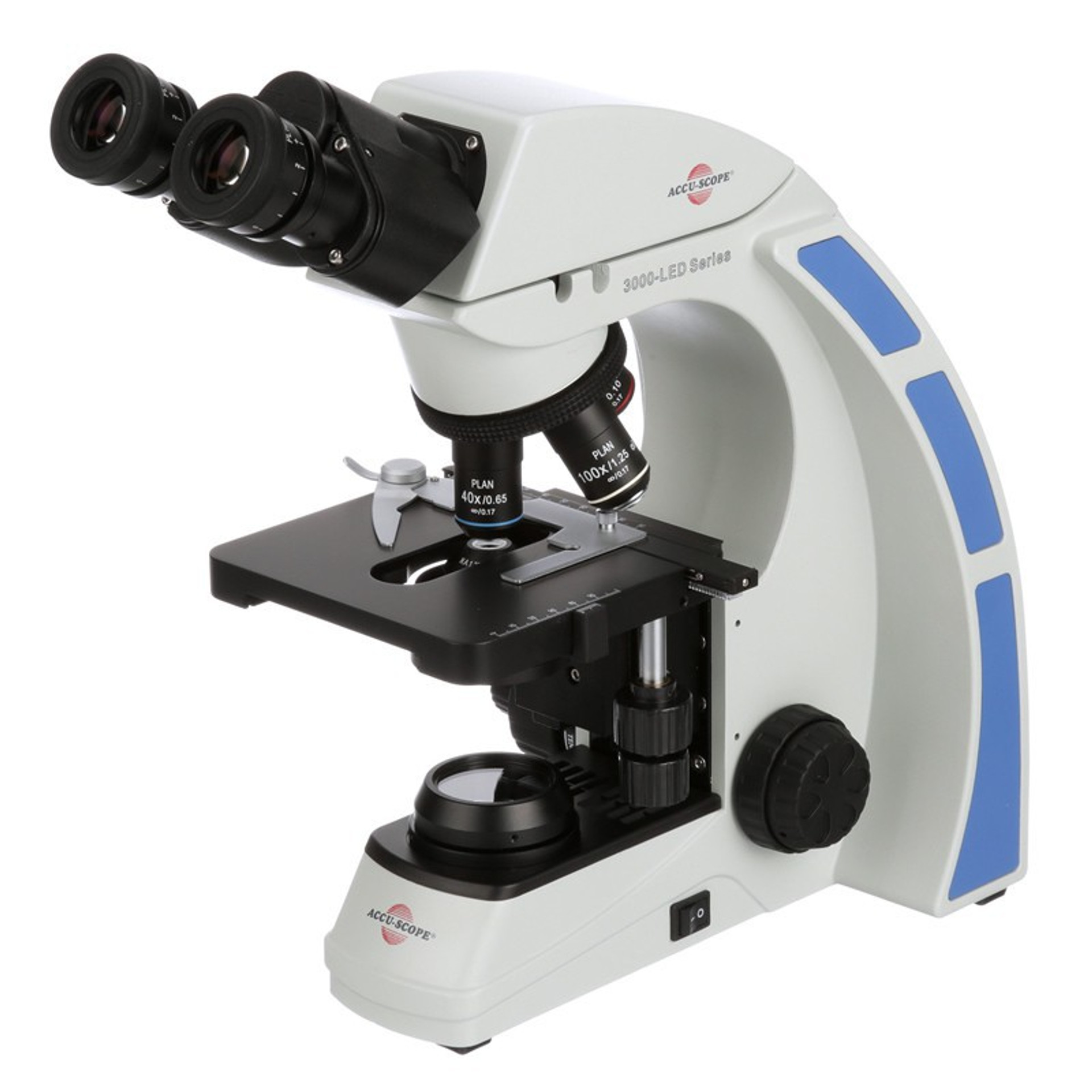
Compound Microscope Parts - Labeled Diagram and their Functions A compound microscope is the most common type of light (optical) microscopes. The term "compound" refers to the microscope having more than one lens. Basically, compound microscopes generate magnified images through an aligned pair of the objective lens and the ocular lens.

Labelled compound microscope
Parts of a microscope with functions and labeled diagram - Microbe Notes Microscopes are instruments that are used in science laboratories to visualize very minute objects such as cells, and microorganisms, giving a contrasting image that is magnified. Microscopes are made up of lenses for magnification, each with its own magnification powers. Compound Microscope Labeled Diagram | Quizlet Compound Microscope Labeled + − Flashcards Learn Test Match Created by meganplocher734 Terms in this set (14) Eyepiece/Ocular lens Contains the ocular lens Body tube A hollow cylinder that holds the eyepiece. Arm Part that supports the microscope. Stage Supports the slide or specimen Coarse adjustment Knob Microscope Parts and Functions With Labeled Diagram and Functions How does a Compound Microscope Work? Before exploring microscope parts and functions, you should probably understand that the compound light microscope is more complicated than just a microscope with more than one lens.
Labelled compound microscope. A Study of the Microscope and its Functions With a Labeled Diagram ... These labeled microscope diagrams and the functions of its various parts, attempt to simplify the microscope for you. However, as the saying goes, 'practice makes perfect', here is a blank compound microscope diagram and blank electron microscope diagram to label. Compound Microscope: Parts of Compound Microscope - BYJUS A compound microscope is used for viewing samples at high magnification (40 - 1000x). Further Reading Microbiology Early Experiments On Photosynthesis Test Your Knowledge On Study Of The Parts Of A Compound Microscope! Put your understanding of this concept to test by answering a few MCQs. Click 'Start Quiz' to begin! Compound Microscope- Definition, Labeled Diagram, Principle, Parts, Uses A compound light microscope mostly uses a low voltage bulb as an illuminator. The stage is the flat platform where the slide is placed. Nosepiece and Aperture Nosepiece is a rotating turret that holds the objective lenses. The viewer spins the nosepiece to select different objective lenses. Compound Microscope Parts - Labeled Diagram and their Functions (2023) The term "compound" refers to the microscope having more than one lens. Basically, compound microscopes generate magnified images through an aligned pair of the objective lens and the ocular lens. In contrast, "simple microscopes" have only one convex lens and function more like glass magnifiers. [In this figure] Two "antique ...
Labelled Diagram of Compound Microscope Labelled Diagram of Compound Microscope Article Shared by ADVERTISEMENTS: The below mentioned article provides a labelled diagram of compound microscope. Part # 1. The Stand: The stand is made up of a heavy foot which carries a curved inclinable limb or arm bearing the body tube. Compound Microscope - Types, Parts, Diagram, Functions and Uses Meaning. Simple microscope - It is a convex lens of small focal length and its primary use is to see a magnified image of small objects. Compound microscope - It is an optical instrument consists of two convex lenses of short focal lengths primarily used for observing a highly magnified image of minute objects. Lenses. Diagram of a Compound Microscope - Biology Discussion 1. It is noted first that which objective lens is in use on the microscope. 2. Stage micrometer is positioned in such a way that it is in the field of view. 3. The eyepiece is rotated so that the two scales, the eyepiece or ocular scale and the stage micrometer scale, are parallel. 4. (a) Draw a labelled ray diagram of a compound microscope. (b) Derive an ... (a) Labelled diagram of compound microscope. The objective lens form image A' B' near the first focal point of eyepiece. (b) Angular magnification of objective lens m 0 = linear magnification h'/h. where L is the distance between second focal point of the objective and first focal point of eyepiece.If the final image A'' B'' is formed at the near point.
Microscopy: Intro to microscopes & how they work (article) - Khan Academy In most cases, the part of a cell or tissue that we want to look at isn't naturally fluorescent, and instead must be labeled with a fluorescent dye or tag before it goes on the microscope. The leaf picture at the start of the article was taken using a specialized kind of fluorescence microscopy called confocal microscopy. Compound Microscope Parts A high power or compound microscope achieves higher levels of magnification than a stereo or low power microscope. It is used to view smaller specimens such as cell structures which cannot be seen at lower levels of magnification. Essentially, a compound microscope consists of structural and optical components. Compound Microscope: Definition, Diagram, Parts, Uses, Working ... - BYJUS The compound microscope is mainly used for studying the structural details of cell, tissue, or sections of organs. The parts of a compound microscope can be classified into two: Non-optical parts Optical parts Non-optical parts Base The base is also known as the foot which is either U or horseshoe-shaped. (i) Draw a neat labelled ray diagram of a compound microscope. Explain ... Working: Suppose a small object AB is placed slightly away from the first focus F 0 ' of the objective lens. The objective lens forms the real, inverted and magnified image A'B', which acts as an object for eyepiece. The eyepiece is so adjusted that the image A'B' lies between the first focus F e ' and the eyepiece E. The eyepiece forms its image A'' B'' which is virtual, erect and magnified.
Compound Light Microscope: Everything You Need to Know A compound microscope is a type of light microscope that uses a compound lens system to magnify specimens for up to 1000x or more. It's made up of at least one objective lens and at least one ocular lens, as well as a light source, condenser, and other essential parts.
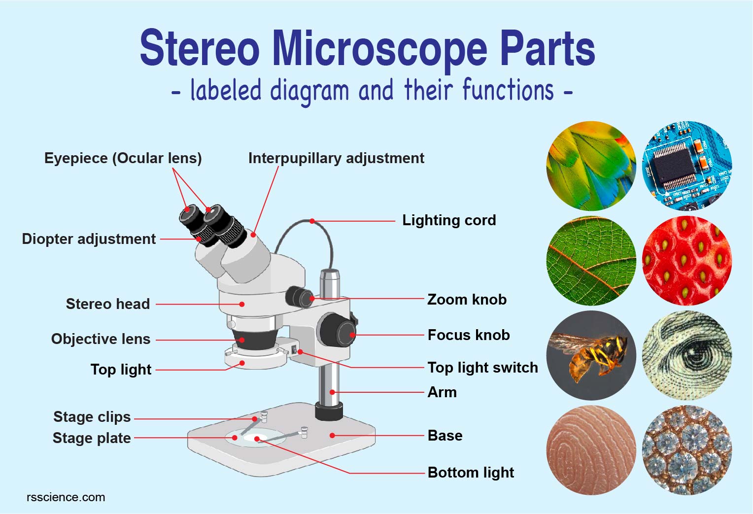
Parts of Stereo Microscope (Dissecting microscope) - labeled diagram ... Unlike a compound microscope that can only see a very thin specimen, stereo microscopes can be used for viewing almost anything you can fit under them. However, stereo microscopes offer lower magnification, typically 5x-50x, comparing to compound microscopes. Below is an example showing the difference between viewing with compound vs stereo ...
Label the microscope — Science Learning Hub Label the microscope Interactive Add to collection Use this interactive to identify and label the main parts of a microscope. Drag and drop the text labels onto the microscope diagram. diaphragm or iris base eye piece lens fine focus adjustment light source stage coarse focus adjustment high-power objective Download Exercise
Light microscopes - Cell structure - Edexcel - BBC Bitesize The compound microscope uses two lenses to magnify the specimen: the eyepiece and an objective lens. In most microscopes, there is a choice of objectives to use. Magnification can therefore...
Parts of the Microscope with Labeling (also Free Printouts) 5. Knobs (fine and coarse) By adjusting the knob, you can adjust the focus of the microscope. The majority of the microscope models today have the knobs mounted on the same part of the device. Image 5: The circled parts of the microscope are the fine and coarse adjustment knobs. Picture Source: bp.blogspot.com.
Microscope Parts and Functions With Labeled Diagram and Functions How does a Compound Microscope Work? Before exploring microscope parts and functions, you should probably understand that the compound light microscope is more complicated than just a microscope with more than one lens.
Compound Microscope Labeled Diagram | Quizlet Compound Microscope Labeled + − Flashcards Learn Test Match Created by meganplocher734 Terms in this set (14) Eyepiece/Ocular lens Contains the ocular lens Body tube A hollow cylinder that holds the eyepiece. Arm Part that supports the microscope. Stage Supports the slide or specimen Coarse adjustment Knob
Parts of a microscope with functions and labeled diagram - Microbe Notes Microscopes are instruments that are used in science laboratories to visualize very minute objects such as cells, and microorganisms, giving a contrasting image that is magnified. Microscopes are made up of lenses for magnification, each with its own magnification powers.












Post a Comment for "40 labelled compound microscope"