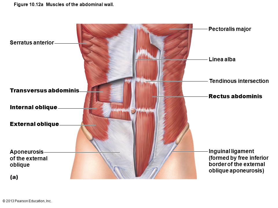
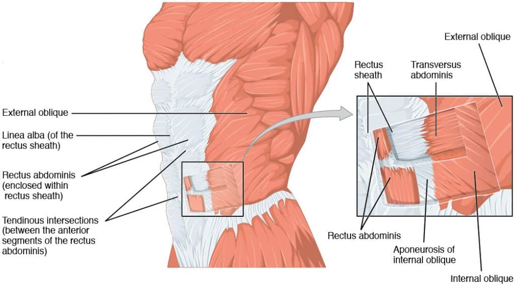
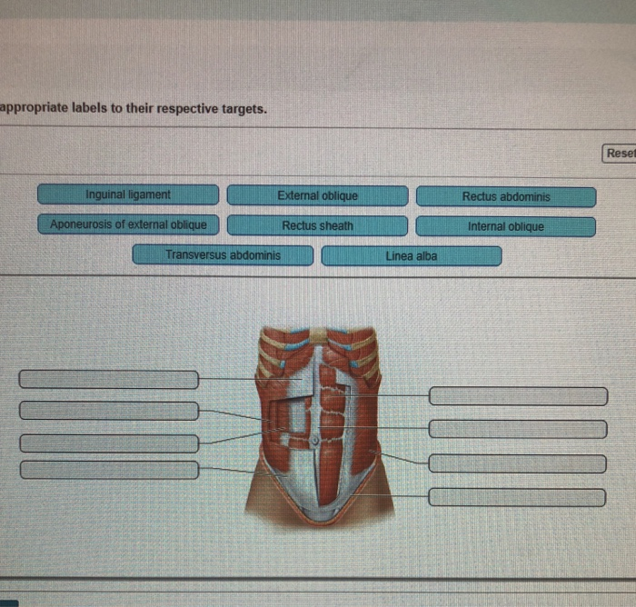
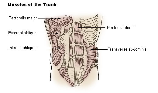
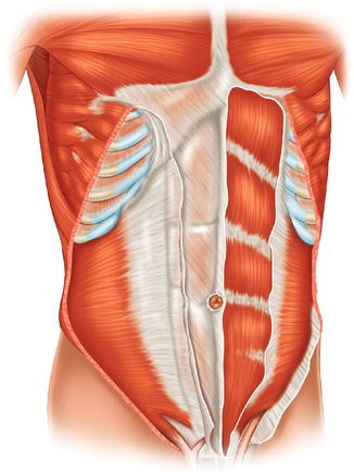
41 label the muscles of the abdominal wall in the figure
11.4.pdf - 11.4 Muscles of the Abdominal Wall Learning... The anterolateral wall of the abdomen is reinforced by four pairs of muscles that collectively compress and hold the abdominal organs in place: the external oblique, internal oblique, transversus abdominis, and rectus abdominis(figure 11.14). These muscles also work together to flex and stabilize the vertebral column. Anatomy, Abdomen and Pelvis, Abdominal Wall - NCBI Bookshelf The abdomen describes a portion of the trunk connecting the thorax and pelvis. An abdominal wall formed of skin, fascia, and muscle encases the abdominal cavity and viscera. The abdominal wall does not only contain and protect the intra-abdominal organs but can distend, generate intrabdominal pressure, and move the vertebral column. Detailed knowledge of the components of the abdominal wall is ...
Chapter 11 and 12 Flashcards | Quizlet True Label the muscles that move the glenohumeral joint in the figure. Hold your arm out so that it is completely extended. The triceps brachii is causing the extension, so it is acting as what? Agonist

Label the muscles of the abdominal wall in the figure
Abdominal wall: Layers, muscles and fascia | Kenhub Lateral flat muscle group situated on either side of the abdomen, which includes three muscles: external oblique, internal oblique and transversus abdominis . Anterior vertical muscles situated bilaterally to the median fibrous structure called linea alba. They are called rectus abdominis and pyramidalis muscles. Solved FIGURE 24.6 Label the muscles of the abdominal wall. | Chegg.com Expert Answer Answer- Labelling of muscles of the abdominal wall. ( Also represented in the image provided below ) External oblique muscle Internal oblique muscle Rectus abdominis muscle Transverse abdominis muscle Name the abdominal muscles a surgeon would incise … View the full answer PDF Document1 - Gore's Anatomy & Physiology 4. part of the abdominal girdle: forms the external lateral walls of the abdomen 5. Acting alone, each muscle of this pair rums the head toward the opposite shoulder 6. and 7. Besides the two abdominal muscles (pairs) named above. two pairs that help form the natural abdominal girdle 8_ Deep muscles of the thorax that promote
Label the muscles of the abdominal wall in the figure. Ch. 15 Cardiovascular Flashcards | Quizlet When _____ muscles contract, they massage the veins, pushing the blood _____ the heart. skeletal; toward ... Determine whether each term describes a part of the heart wall or the part of the coverings of the heart. Then place each label in the appropriate category. ... The figure demonstrates the effects of skeletal muscle contraction on venous ... (Get Answer) - FIGURE 24.6 Label the muscles of the abdominal wall ... FIGURE 24.6 Label the muscles of the abdominal wall. APIR Pectoralis minor Internal intercostals... FIGURE 24.6 Label the muscles of the abdominal wall. APIR Pectoralis minor Internal intercostals Serratus anterior - External intercostals Rectus sheath- . Rectus abdominis External oblique Inguinal ligament Nov 16 2021 08:34 AM Expert's Answer label the muscles of the abdominal wall Quiz - PurposeGames.com This is an online quiz called label the muscles of the abdominal wall There is a printable worksheet available for download here so you can take the quiz with pen and paper. Your Skills & Rank Total Points 0 Get started! Today's Rank -- 0 Today 's Points One of us! Game Points 10 You need to get 100% to score the 10 points available Muscles of the trunk: Anatomy, diagram, pictures | Kenhub Pyramidalis is a variable muscle of the abdominal wall, being absent in about 20% of the population. Abdominal oblique muscles It's time to take a look at the three flat muscles of the anterolateral abdominal wall. The first two are the abdominal oblique muscles. These include the external abdominal oblique and the internal oblique muscles.
Label the muscle and other structures indicated in - Course Hero inspiratory expiratory muscles of the rib cage wall sternocleidomastoid scalenus anterior, medius, and posterior pectoralis major pectoralis minor subclavius serratus anterior external intercostal internal intercostal (between ribs) internal intercostal (between costal cartilages) transversus thoracis latissimus dorsi serratus posterior superior … Axial Muscles of the Abdominal Wall and Thorax - Course Hero There are four pairs of abdominal muscles that cover the anterior and lateral abdominal region and meet at the anterior midline. These muscles of the anterolateral abdominal wall can be divided into four groups: the external obliques, the internal obliques, the transversus abdominis, and the rectus abdominis (Figure 1, Figure 2, and Table 1). Anatomy Muscle Quiz- Trunk & Abdominal - ProProfs Quiz Create your own Quiz. The muscles of the trunk include those that move the vertebral column, the muscles that form the thoracic and abdominal walls, and those that cover the pelvic outlet. Test just how much you know about these muscles by taking the simple quiz below. All the best and share it with your friends! Axial Muscles of the Abdominal Wall and Thorax - Course Hero There are four pairs of abdominal muscles that cover the anterior and lateral abdominal region and meet at the anterior midline. These muscles of the anterolateral abdominal wall can be divided into four groups: the external obliques, the internal obliques, the transversus abdominis, and the rectus abdominis (Figure 16.16 and Table 16.6).
abdomen anatomy muscles Axial Muscles Of The Abdominal Wall And Thorax | Anatomy And Physiology I courses.lumenlearning.com. muscles abdominal abdomen lumbar posterior anterior anatomy axial thorax spine movements assist lower move figure. Abdominal Muscle Labeling Quiz . abdominal muscle quiz. Muscles Of Abdomen - YouTube The Anterolateral Abdominal Wall - Muscles - TeachMeAnatomy Assists in forceful expiration by pushing the abdominal viscera upwards. Is involved in any action (coughing, vomiting, defecation) that increases intra-abdominal pressure. The anterolateral abdominal wall consists of four main layers (external to internal): skin, superficial fascia, muscles and associated fascia, and parietal peritoneum. Muscle Lab 22 Figure 22.5 Muscle of Abdominal Wall - Quizlet Muscle Lab 22 Figure 22.5 Muscle of Abdominal Wall STUDY Learn Write Test PLAY Match + − Created by THagge TEACHER Terms in this set (4) External Oblique ... Rectus Abdominis ... Internal Oblique ... Transversus Abdominis ... OTHER SETS BY THIS CREATOR Word Parts Quiz 13 10 terms THagge TEACHER Word Parts 12 10 terms THagge TEACHER Muscle Sarcomere Solved FIGURE 24.6 Label the muscles of the abdominal wall. - Chegg Expert Answer Labelling of muscles of the abdominal wall. ( Also represented in the image provided below ) External oblique muscle Internal oblique muscle Rectus abdominis muscle Transverse abdominis muscle Name the abdominal muscles a surgeon would incise from su … View the full answer
Muscles of the Deep Back, Abdominal Wall, and Pelvic ... complete parts a and b. 1 figure 22.1 label the three deep back muscle groups of the erector spinae group.use the following options: iliocostalis (lateral group), longissimus (intermediategroup), spinalis (medial group).figure 22.2 label the muscles of the abdominal wall. 2 figure 22.3 label the muscles of the male pelvic outlet.figure 22.4 …
Figure 10.12 (a): Muscles of the abdominal wall - Quizlet Rectus abdominis Flex and rotate lumbar region of vertebral column Transversus abdominis Compresses abdominal contents Internal oblique Flex vertebral column and compress abdominal wall, aid muscles of back in rotating trunk and flexing laterally (same as external oblique) External oblique
Axial Muscles of the Abdominal Wall and Thorax The muscles of the vertebral column, thorax, and abdominal wall extend, flex, and stabilize different parts of the body's trunk. The deep muscles of the core of the body help maintain posture as well as carry out other functions. The brain sends out electrical impulses to these various muscle groups to control posture by alternate contraction ...
11.5 Muscles of the Pectoral Girdle and Upper Limbs Figure 11.23 Muscles That Move the Humerus (a, c) The muscles that move the humerus anteriorly are generally located on the anterior side of the body and originate from the sternum (e.g., pectoralis major) or the anterior side of the scapula (e.g., subscapularis). (b) The muscles that move the humerus superiorly generally originate from the ...
Solved FIGURE 24.6 Label the muscles of the abdominal wall - Chegg Transcribed image text: FIGURE 24.6 Label the muscles of the abdominal wall MPIRE Pectorate or -Intera intercostal Sawatusan External intercosta Rectus et Ingunnalligament CRITICAL THINKING ASSESSMENT Name the abdominal muscles a surgeon would incise from superficial to deep when performing an appendectomy Siniz omul zamboup
Lab 8: Anterolateral abdominal wall and inguinal region - ESFCOM Learning LAB OUTLINE. 1 Identify the layers of fascia in the anterolateral abdominal wall. 2 Identify the external oblique, internal oblique, and transversus abdominis muscles and discuss attachments and functions. 3 Identify the rectus sheath and the rectus abdominis muscle and discuss attachments and functions. 4 Identify the nerves and vessels supplying the abdominal wall
Muscle Lab 22: Figure 22.1 Muscles of the Abdominal Wall Only $35.99/year Muscle Lab 22: Figure 22.1 Muscles of the Abdominal Wall STUDY Learn Write Test PLAY Match Created by THaggeTEACHER Terms in this set (7) External oblique Internal oblique Transversus abdominis Rectus abdominis Linea Alba Unbilicus Aponeurosis Sets found in the same folder Muscle Lab 23 Figure 23.2 Muscles of the… 6 terms
Quantitative Anatomical Labeling of the Anterior Abdominal Wall These three muscles are connected superiorly to the 5th through 12th ribs, inferiorly to the iliac crest, and extend across the anterior abdominal wall toward the rectus muscle along the midline. They form the lateral barriers of the abdominal wall. On CT, these three muscles can be identified as a singular muscular group.
Muscles_of_the_Abdominal_Wall__Pelvic_Outlet.pdf Theanteriorand lateralwalls ofthe abdomencon- tain broad, flattened muscles arranged in layers. Thesemusclesconnecttherib cageand vertebralcol- umntothe pelvicgirdle.Theabdominalwallmuscles compress the abdominal visceralorgans, help main- tainposture,assist inforcefulexhalation,andcontrib- utein trunkflexion andwaistrotation.
Abdominal Muscles: Anatomy and Function - Cleveland Clinic Abdominal Muscles. Your abdominal muscles have many important functions, from holding organs in place to supporting your body during movement. There are five main muscles: pyramidalis, rectus abdominus, external obliques, internal obliques, and transversus abdominis. Ab strains and hernias are common, but several strategies can keep your abs ...
chapter 11 a&p Flashcards | Quizlet label the muscles of the abdominal wall in the figure left -rectus abdominis right -transverse abdominis -internal oblique -external oblique classify the actions with the muscles of the abdominal wall that perform them. some answers will be used more than once. obliques -lateral flexion of vertebral column -compresses abdominal wall
Sarcoidosis - Wikipedia Sarcoidosis is a systemic inflammatory disease that can affect any organ, although it can be asymptomatic and is discovered by accident in about 5% of cases. Common symptoms, which tend to be vague, include fatigue (unrelieved by sleep; occurs in up to 85% of cases ), lack of energy, weight loss, joint aches and pains (which occur in about 70% of cases), arthritis …
PDF Document1 - Gore's Anatomy & Physiology 4. part of the abdominal girdle: forms the external lateral walls of the abdomen 5. Acting alone, each muscle of this pair rums the head toward the opposite shoulder 6. and 7. Besides the two abdominal muscles (pairs) named above. two pairs that help form the natural abdominal girdle 8_ Deep muscles of the thorax that promote
Solved FIGURE 24.6 Label the muscles of the abdominal wall. | Chegg.com Expert Answer Answer- Labelling of muscles of the abdominal wall. ( Also represented in the image provided below ) External oblique muscle Internal oblique muscle Rectus abdominis muscle Transverse abdominis muscle Name the abdominal muscles a surgeon would incise … View the full answer
Abdominal wall: Layers, muscles and fascia | Kenhub Lateral flat muscle group situated on either side of the abdomen, which includes three muscles: external oblique, internal oblique and transversus abdominis . Anterior vertical muscles situated bilaterally to the median fibrous structure called linea alba. They are called rectus abdominis and pyramidalis muscles.
:background_color(FFFFFF):format(jpeg)/images/library/12218/ventral-trunk-muscles_english__1_.jpg)



:watermark(/images/watermark_only_sm.png,0,0,0):watermark(/images/logo_url_sm.png,-10,-10,0):format(jpeg)/images/anatomy_term/ilioinguinal-nerve/LrjoPagBvWY81UExQQ7lww_Nervus_ilioinguinalis_02.png)










Post a Comment for "41 label the muscles of the abdominal wall in the figure"