42 labeled histology slides
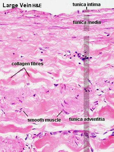
Histology guide: Definition and slides | Kenhub At a histological level, both the heart and blood vessels consist of three layers: Endothelial layer - epithelial tissue formed by simple squamous (endothelial) cells. In the heart, this layer is referred to as endocardium. Muscular layer - smooth muscle in the blood vessels, cardiac muscle (myocardium) in the heart. Histology of testes & epididymis - SlideShare 25. epididymis 1. Highly coiled mucular tubule 2. 6 meter long, made up of smooth muscle 3. smooth muscle arranged in circular pattern 4. is lined by pseudostratified columnar epithelium with stereocilia. [non- motile cilia] 5. Can store sperms for several months 6. Continues as ductus (vas) deferens smooth muscle pseudostratifi ed columnar ...
Online Histology Made Easy Slides Atlas | MedicForYou 21 LS of thin skin Histology slide 22 Lungs Histology slide 23 Lymph node Histology slide 24 Mixed gland Histology slide 25 Mucus glands Histology slide 26 Muscular artery Histology slide 27 Esophagus Histology slide 28 Ovary Histology slide 29 Palatine tonsil Histology slide 30 Pituitary gland Histology slide 31 Prostate Histology slide

Labeled histology slides
Histology Page - Napa Valley College Digestive Histology. glands in the tongue 40X. muscle in the tongue 40X. stratified squamous epith tongue 40X.bmp. taste buds 40X.bmp. tastebud in the tongue 400X. duct salivary gland 400X. submandibular salivary gland 40X. serous acini salivary gland 400X. Ovary histology slide diagram and identification points In the ovary histology slide labeled diagram, you will find all the features that are mentioned above. Identification of other slides from the female organs Uterine tube histology slide with its identification points Uterus histology slide with its identification points Mammary glands histology slide with its identification points Colon Histology Slide with Labeled Diagram - AnatomyLearner Colon Histology Slide with Labeled Diagram 04/06/2022 04/06/2022 by anatomylearner The colon histology slide possesses the typical four layers of a tubular organ - mucosa, submucosa, muscularis, and serosa. But, there are no permanent plica circularis and villi in the colon slide as found in the different segments of the small intestine.
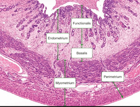
Labeled histology slides. Slides of Histology | Anatomy and Physiology I | | Course Hero Slides of Histology Learning Objectives Be able to describe the functions of cells commonly found in connective tissue and identify them. Be able to recognize interstitial (fibrillar) collagens and elastic fibers at the light and electron microscopic levels. PPT - Histology Slides for the Respiratory System PowerPoint ... Chart and Diagram Slides for PowerPoint - Beautifully designed chart and diagram s for PowerPoint with visually stunning graphics and animation effects. Our new CrystalGraphics Chart and Diagram Slides for PowerPoint is a collection of over 1000 impressively designed data-driven chart and editable diagram s guaranteed to impress any audience. Uterus histology slide with labeled diagram and identification points Uterus histology slide labeled diagram The uterine cavity is lined by simple columnar epithelium (covering of the mucosa layers) Presence of highly cellular endometrium that contains the uterine glands There is thick myometrium having a thick middle circular layer of smooth muscle with many blood vessels. Histology Guide - virtual microscopy laboratory Histology Guide solves this problem by recreating the look and feel of a microscope in an intuitive, browser-based interface. An Aperio slide scanner was used to obtain a high-resolution image of each slide in its entirety. Large tissues are up to 34 GB for a single, uncompressed image of 150,000 x 75,000 pixels.
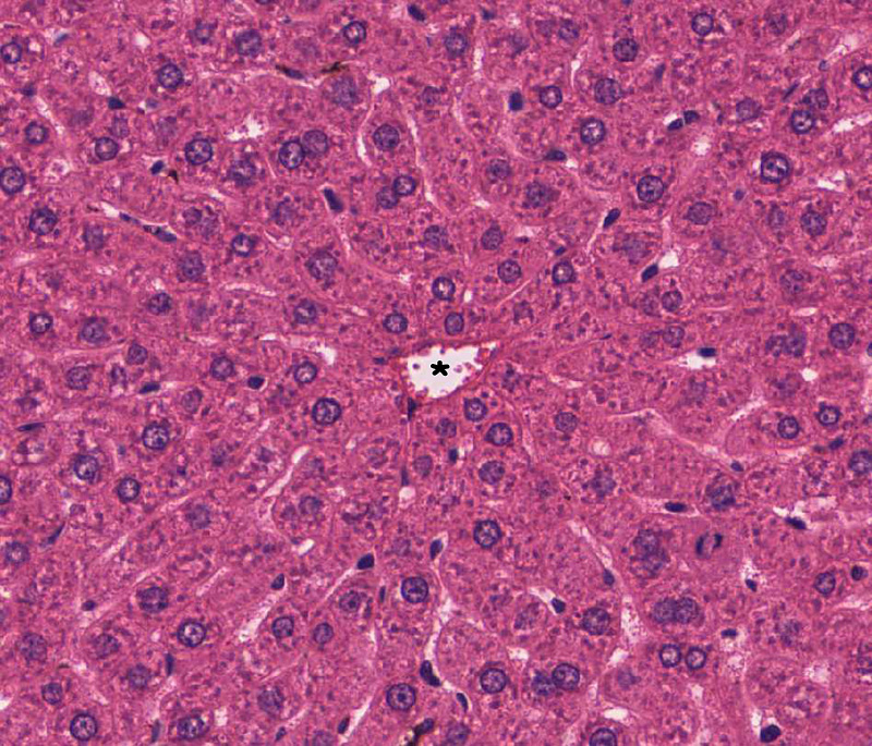
Histology Slides 1 - Loyola University Chicago Esophagus Histology Slides Labeled - esophagus histology esophagus ... Here are a number of highest rated Esophagus Histology Slides Labeled pictures upon internet. We identified it from reliable source. Its submitted by direction in the best field. We admit this kind of Esophagus Histology Slides Labeled graphic could possibly be the most trending subject subsequently we ration it in google improvement or facebook. Histology of the Liver- - SlideShare The Liver • Largest gland of the body. • 1500 grams and 2.5% of total body weight. • Location: - Right hypochondrium - Epigastric region - Left hypochondrium. 3. Vascular Supply of the Liver • Receives dual vascular supply: -Hepatic Portal Vein (75%) - Hepatic Artery (25%) • Both vessels enter the liver via Porta hepatis. 4. Histology Slides Identification | Unmsa What slide is this? Small intestine Oviduct Ureter Large intestine 22. What slide is this? Smooth cartilage Elastic cartilage Fibrocartilage Hyaline cartilage 23. What is the dimension of the slide above 8cm by 5cm by 3cm 17cm by 15cm by 12cm 12cm by 6cm by 3cm 45cm by 22cm by 18cm 24. What slide is this? Thick Muscularis Vas deferns Testis Penis
Microscope Slides of Cells and Tissues | Histology Guide This virtual slide box contains 275 microscope slides for the learning histology. Fig 023 Types of Tissue Cells and Tissues Tissues are classified into four basic types: epithelium, connective tissue (includes cartilage, bone and blood), muscle, and nervous tissue. Chapter 1 The Cell Chapter 2 Epithelium Chapter 3 Connective Tissue Chapter 4 Muscle Histology: Labelled Slides | SchoolWorkHelper Histology: Labelled Slides Aorta Basophils Cardiac Muscle Cardiac Muscle Longtudinal Cerebellar Cortex Cerebral Cortex Spinal Cord Eosinophils Epiphyseal plate Femoral Artery Femoral Vein Howship's lacunae Jejunum lamina propria Liver Lymph node Lymphocyte Monocyte Spinal cord (silver) Neutrophil Pacinian corpuscle Peripheral Nerve Histology Slides - Napa Valley College BIOL 218 Human Anatomy; BIOL 219 Human Physiology; BIOL 240 General Zoology; People Sites > Dan Clemens > Histology Slides > > People ... Histology Slides Clemens BIOL 218 Histology Slide Images Exercise 2 - Cells 02_Blood_100X 02_Blood_400X 02_Neurons ... Anatomy Department donates Histology slides | ANATOMY Anatomy Department donates Histology slides The Department of Anatomy has been responsible for teaching histology to medical students since the inception of the UCSF School of Medicine. Over the years, we have prepared a very large collection of glass microscope slides, which are primarily of human tissues.
Basic Histology -- Cuboidal Epithelium, Two Layers | Histology slides ...
PPT Histology Slides - Oakton Community College Histology Slides Biology 131 - Anatomy and Physiology Instructor: Ruth Williams Epithelial Tissues No intercellular matrix. Avascular Contains nerve endings Lie on a basement membrane. Able to undergo mitosis. Develop from all three fetal tissues. One surface of cells is exposed to a space or cavity.

Cartilage Histology - Hyaline cartilage - histology slide - | Histology ...
Histology Slides Identification from Different Organ Systems This article will show you histology slides from the following different organs system of an animal's body with identifying features. #1. Histology slide of epithelial tissue #2. General connective tissue histology slide #3. Histology slides of special connective tissue (blood, bone, and cartilage) #4. Muscular tissue histology slide #5.
Liver, Gallbladder and Pancreas | histology
histology human anatomy tissue slides Flashcards and Study Sets - Quizlet epithelial tissue histology slides stratified squamous epithelium simple ciliated columnar epithelium pseudostratified ciliated columnar epit… flattened tile-like cells in surface layer, rounder cells in b… elongated cells, oval shaped nuclei, single layer, projections… Actually a single layer of cells of varying height, some not r… 11 sets Kenhub
ANAT2511 Circulatory System - Embryology
Oral histology slides - Anatomicum.com Oral histology slides This page contains a list of labeled histology images taken from the dental education program at the University of Oslo. If you want to help translate these pages and all labels to your language, please mail me at feedback@anatomicum.com. Odontogenesis Bud stage Advanced bell stage Bell stage Bud stage Cap stage Teeth
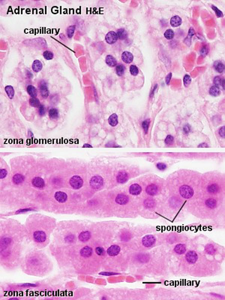
File:Adrenal histology 004.jpg - Embryology
Anatomy and Physiology - Histology Slides Flashcards | Quizlet 21 terms callie_pavlicek8 Anatomy and Physiology - Histology Slides STUDY PLAY Simple Squamous Epithelium Stratified Squamous Epithelium Simple Columnar Epithelium Stratified Columnar Epithelium Pseudostratified Columnar Epithelium Simple Cuboidal Epithelium Stratified Cuboidal Epithelium Transitional Epithelium Areolar loose connective tissue
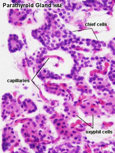
File:Parathyroid histology 002.jpg - Embryology
Histology Slides 1 - Loyola University Chicago Slide 15 EM of liver cell (hepatocyte), showing aggregates of glycogen (1). Compare their size with the ribosomes lining the endoplasmic reticulum (2). Gallbladder Slide 16 Wall of gall bladder, showing high, branching mucosal folds. These are not villi. The rest of the wall contains connective tissue and thin strands of smooth muscle. Slide 17

Histology Slides Database: histological diagram of ovary
Histology Microscope Slides | Carolina.com Study the anatomy of cells and tissues in different plants and animals with histology slides from Carolina! Our prepared anatomy and physiology tissue slides are ready for you to teach and examine right out of the box. Our samples range from animal tissue and cells to human samples. Whether your skill level is on a beginner stage or more ...
Chapter 27: The Reproductive System Flashcards | Easy Notecards
[Lungs Histology Slides Labeled] - 16 images - lab exercises lung ... Here are a number of highest rated Lungs Histology Slides Labeled pictures on internet. We identified it from well-behaved source. Its submitted by organization in the best field. We understand this kind of Lungs Histology Slides Labeled graphic could possibly be the most trending subject in imitation of we ration it in google pro or facebook.
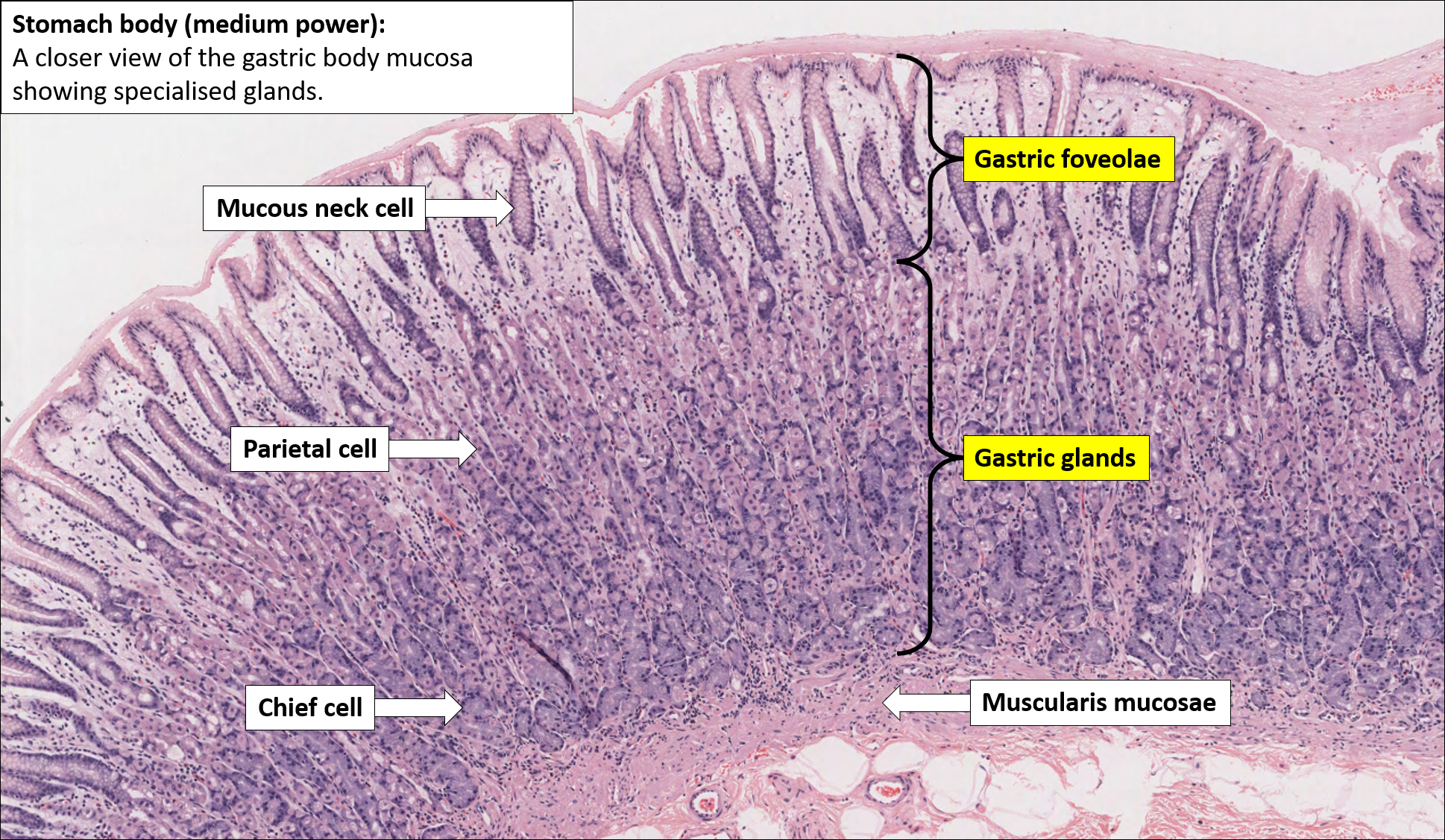
Stomach – Normal Histology – NUS Pathweb
Duke Histology - Slide Index University of Michigan Slide Collection. Below is an index of slides from the University of Michigan Histology collection compiled by Dr. Kent Christensen, Ph.D., J. Matthew Velkey, Ph.D., Lloyd M. Stoolman, M.D., Laura Hessler, and Diedra Mosley-Brower. The ImageScope links requires a Windows machine running ImageScope ( If you need a copy ...
Post a Comment for "42 labeled histology slides"