41 label the diagram of the nucleotides below
Topic 7.3, 7.4, 7.5: DNA Replication, Transcription ... - Brainscape two polymers shown; arranged in a double helix; sugar shown connected to base; sugar-phosphate backbone shown; If only one nucleotide is drawn, award [2 max] sugar identified as deoxyribose; hydrogen bonding between bases shown; diagram shows complementary base pairing / A bonded to T, C with G; Award previous mark if bases (unlabelled) are shown in the diagram but the complementary base ... Sketch And Label A Nucleotide : Solved Problem 1 What Are The Three ... The general structural representation of a nucleotide is shown below. A bond between nucleotides in rna and dna molecules. Draw a nucleotide and label the three main parts. See below the above structure is a color (magenta)nucleotide. Using arrows and brackets, identify and label at least one example of each of the following.
16identify and label the diagram below 17 list the 16.Identify and label the diagram below: 1. Primase joins RNA nucleotides into a primer 2. DNA pol 3 adds DNA nucleotides to the primer, forming Okazaki fragment 1. 3. After reaching the next RNA primer to the right, DNA pol 3 detaches. 4.
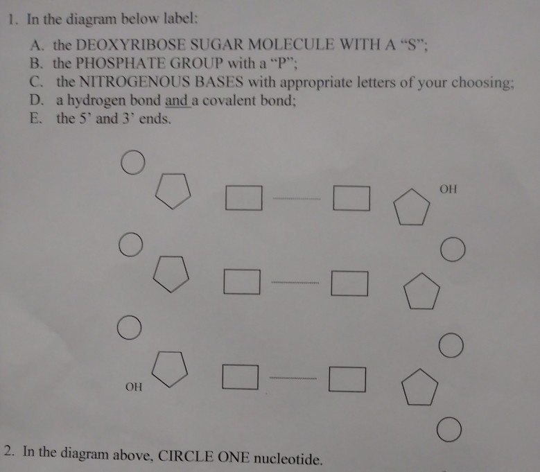
Label the diagram of the nucleotides below
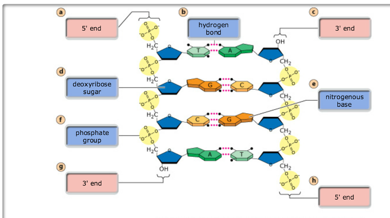
The Structure of DNA This figure is a diagram of a short stretch of a DNA molecule which is unwound and flattened for clarity. The boxed area at the lower left encloses one nucleotide. Each nucleotide is itself make of three subunits: A five carbon sugar called deoxyribose (Labeled S) Draw And Label A Dna Nucleotide Indicating The Position And Number Of ... See below the above structure is a color (magenta)nucleotide. 3.3.5 draw and label a simple diagram of the molecular structure of dna. Draw the general structure of a nucleotide and a nucleoside. Nucleic acids are made up of chains of many repeating units called nucleotides (see bottom left of figure 1 below). Chapter 13 Flashcards | Quizlet The diagram represents DNA that is part of the RNA‑coding sequence of a transcription unit. The bottom strand is the template strand. 5′-GCATATGCGGTAC-3′3′-CGTATACGCCATG-5′5′-GCATATGCGGTAC-3′3′-CGTATACGCCATG-5′ Give the sequence found on the RNA molecule that is transcribed from the above DNA molecule.
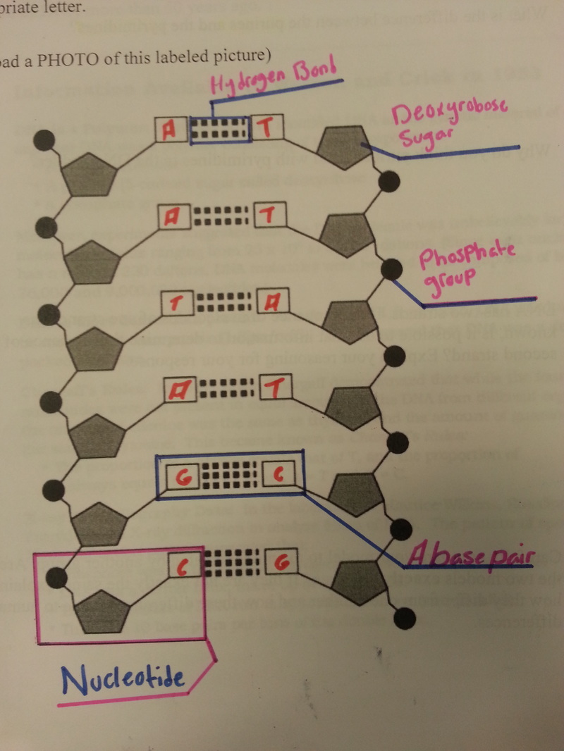
Label the diagram of the nucleotides below. PDF Label the structures of this single nucleotide ... - Weebly Label the structures of this single nucleotide. a. Phosphate b. Deoxyribose sugar c. Nitrogenous base Complete the table below to show the pairings of the bases in DNA: PurinePyramidine Guamine Cytosine Adenine Thymine State where the base uracil can be found. RNA In the space below, draw a single strand of three nucleotides, naming the bonds ... DNA Molecule Label Diagram | Quizlet Molecule found on the side of a DNA molecule Double Helix two strands of nucleotides wound about each other; structure of DNA Thymine the nucleotide that hydrogen bonds with the nucleotide adenine in DNA Adenine the nucleotide that hydrogen bonds with the nucleotide thymine in DNA or with uracil in RNA Guamine DNA Worksheet.docx - Structure of DNA and Replication Directions: Label ... Label the nucleotides (A, T, G, C) in the DNA molecule below: 14. What is the first step in the process of DNA replication. G C A T 2 Helicase comes in and unzips the helix by breaking hydrogen bonds . Helicase comes in and unzips the helix by breaking hydrogen bonds. 15. Which enzyme is responsible for "unzipping" the DNA double helix? Helicase Solved Please answer the questions and label the | Chegg.com Please answer the questions and label the diagram. This is two nucleotides bonded together. This is how the backbone of a strand of DNA is formed. Manipulate this structure so that you can answer the following questions on the diagram below: 1. Indicate on the drawing and give the name of which subunits of the nucleotides are bonded to form
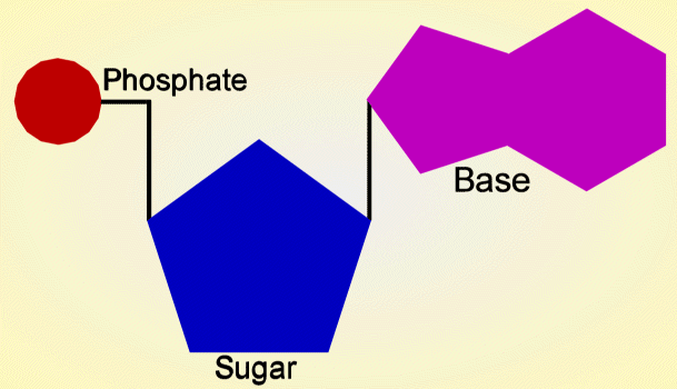
Nucleotide: Structure, Examples and Function - BYJUS Nucleosides are named as Adenosine, Guanosine, Thymidine, Cytidine, Uridine Nucleotide = Nucleoside + Phosphate Nucleotides are named as Adenylic acid, Guanylic acid, Thymidylic acid, Cytidylic acid and Uridylic acid. Nucleotide Structure: DNA Diagram - Science Trends Nucleotides are molecules which serve as the building blocks, or monomer units, for the creation of important polymers like ribonucleic acid or RNA and deoxyribonucleic acid or DNA. As mentioned, nucleotides have three component parts: a five-sided carbon sugar, a nitrogen-containing base, and a phosphate group. (Solved) - Directions: Label the diagram below with the following ... Directions: Label the diagram below with the following choices: • Nucleotide • Deoxyribose • Phosphate group • Base pair • Hydrogen bond • Nitrogenous base 7. A nucleotide is made of three parts: a _____ group, a five carbon _____, and a _____ base. ... What enzyme connects the new nucleotides together and proofreads them? _____ 21. Label the parts of the DNA in the diagram given below. Explain the ... DNA is the hereditary material as it contains genetic information. DNA is a large molecule, consisting of millions of nucleotides. Each nucleotide consists of three compounds. Nucleotides in a DNA (a) A sugar molecule - Deoxy Ribose sugar (b) A nitrogenous base [Purines and Pyrimidines] Purines (Adenine and Guanine) Pyrimidines (Cytosine and ...
Given Below is a Schematic Diagram of a Portion of Dna - Shaalaa.com (a) 2 (b) 2 on each strand (c) 1- Phosphate, 2- Sugar, 3- Nitrogen Base, 4- Hydrogen Bond, 5 - Base (d) Nucleotide Dna Drawing With Labels : Diagrammatic Representation Of ... The dna molecule comprises polymers of nucleotides carrying instructions for development and growth. ... Solved Label The Diagram Below With The Following Labels Anaphase G2 Prepares For Mitosis S Dna Replication Interphase G1 Cell Grows Cytokines Course Hero from The boxed area at the lower left encloses one . 3.3.5 draw and ... Animal Cells: Labelled Diagram, Definitions, and Structure Feb 22, 2022 · They have phospholipid bilayers. There are two types of ER: the rough ER, and the smooth ER. The rough endoplasmic reticulum is rough because it has ribosomes (which is explained below) attached to it. It helps in the synthesis and packaging of proteins. The smooth endoplasmic reticulum doesn’t have ribosomes attached. Dna Replication Label The Diagram - Drawing Diagram Dna and replication worksheet answers label the diagram. DNA polymerase adds nucleotides 5 to 3 Replication fork is formed. Diagram and Label a section of DNA in the box to the right. Helicase opens the DNA and replication forks are formed. The sketch below shows the double helix of DNA.
Solved: 1. In The Diagram Below Label: A. The DEOXYRIBOSE ... | Chegg.com
Dna Diagrams Label - Drawing Diagram DNA Structure Be able to label the following. For each of the circles below DETERMINE if the end is 5 or 3 and label them in the diagram. Structure of the DNA Although DNA is often found as a single - stranded polynucleotide it assumes it s most stable form when double stranded. Label the introns in the. Deoxyribose Nucleic Acid DNA.
DNA Model
PDF Directions: Label the diagram below with the following choices ... Directions: Label the diagram below with the following choices: 7. What is the first step in the process of DNA replication? ... Which enzyme is responsible for facilitating the hydrogen bonding between nucleotides in a new DNA molecule? _____ _____ 10. Which enzyme is responsible for creating the covalent bonds that connect the sugar-phosphate ...
Print Exam 3: Chs. 5 (DNA Structure and Replication Machinery) & 16 ...
(Solved) - Directions: Label the diagram below with the following ... A nucleotide is made of three parts: a group, a five carbon anda base. In a single strand of DNA, the phosphate group binds to the of the next group. Chargaff's rule states that the DNA of any species contains equal amounts of and also equal amounts of In DNA, thymine is complementary to (or pairs with) ;; Cytosine is complementary to 11.

How did scientists find out the composition of DNA? : askscience
Solved ta Directions: Label the diagram below with the | Chegg.com In DNA, guanine always forms. Question: ta Directions: Label the diagram below with the following choices: • Nucleotide • Deoxyribose • Phosphate group • Base pair • Hydrogen bond • Nitrogenous base ca sh fn DNA Molecule: Two Views 2. H H-C H- HE HC -H- -H SHO 5. THO 3. Directions: Complete each sentence.
DNA - Principle of Biomedical Science
Dna Model Drawing With Labels - Solved 1 Label The Structure Of Dna Dna ... One idea would be to look up a labeled diagram of dna and label your own . Nucleic acids are made up of chains of many repeating units called nucleotides (see bottom left of figure 1 below). Learn vocabulary, terms, and more with flashcards, games, and other study tools. ... (see bottom left of figure 1 below). This diagram misses out the ...

30 Draw And Label The Three Parts Of A Nucleotide - Labels Database 2020
GRADE 12 LIFE SCIENCES LEARNER NOTES - Mail & Guardian 3.1 Label the molecules indicated by 2 and 3. (2) 3.2 Using the letters of the genetic code, write down the complementary nitrogenous bases on strand 1 of the DNA double helix, starting from the top. (3) (Remember: A=T/U and G=C) 3.3 Use the table below to determine which three amino acids in the diagram are represented by 4, 5 and 6. (3 x 2) (6)

2. 19: Glucose and ATP - Biology LibreTexts
How do you draw a nucleotide and label its three basic parts? Explanation: The above structure is a nucleotide. It consists of a: phosphate group. 5-carbon sugar, and. nitrogenous base.

Glucose and ATP | CK-12 Foundation
DNA Structure - YouTube Learn about the structure of DNA and how to recognize all the parts in this video!

32 Dna Label - Labels For Your Ideas
Matrix-assisted laser desorption/ionization - Wikipedia In 2015 successful laser post-ionization was reported, using a modified MALDI source operated at an elevated pressure of ~3 mbar coupled to an orthogonal time-of-flight mass analyzer, and employing a wavelength-tunable post-ionization laser, operated at wavelength from 260 nm to 280 nm, below the two-photon ionization threshold of the matrices ...
Post a Comment for "41 label the diagram of the nucleotides below"